INTRODUCTION
Passive fit is one of the most important features to ensure a long-term stability of implant-supported prostheses, since a misfit may result in mechanical and biological failures or even implant loss1. Thus, an accurate transfer from oral cavity to the cast is the key to obtain the clinical success2,3.
The gypsum cast obtained by means of conventional impression is considered appropriate for laboratorial procedures, provided that the position and angulation of dental implants are accurately determined4. However, several factors may influence the final result of dental models manufacturing. Although studies reported a higher accuracy for splinting technique, displacement of components and dimensional changes of impression material may occur during the stone cast fabrication5,6.
CAD-CAM techniques allows the transfer of dental implants in a faster and simple way in comparison with conventional procedures7-9. By means of digital impression, virtual models are generated and enable not only the virtual design of restorations, but also the milling of custom abutments and implant-supported prostheses10. In specific situations on which a whole digital workflow is required, a milled or printed model is fabricated, which allows checking the occlusal relationship, proximal contacts, marginal fit and shape from restoration before try-in in patient`s mouth8,11,12,13.
These models present a higher durability and resistance to alterations in comparison with stone casts7,9,12. However, their accuracy relies on the quality of scanning, which, in turn, is still controversial14-16. Furthermore, geometric defects may also be introduced on the final model due to the manufacturing process, resulting in discrepancies with regard to the implants position transference11,17. Lee and cols., 201511, showed a discrepancy in implant position of milled models due to the manual implant positioning process.
In case of an inaccurate model, the use of a verification jig was preconized to verify and correct a milled polyurethane cast of edentulous patients6,18-21. For the fabrication of partial fixed-dental prosthesis, however, the use of verification jig was not yet assessed. The present study proposed the use of a verification jig to correct the implant position in a partial dentate 3D printed model. The accuracy of the technique is assessed and compared with the conventional impression.
Materials and methods
Master model
An partial dentate maxillary model was made with epoxy resin and used as master model (MM). Dental implants 3.5x8.5 mm (SW Morse, S.I.N. Implant System, São Paulo, Brazil) were installed at the sites of lateral incisor (22), pre-molar (24) and molar teeth (26). Impressions (n=10) were taken from the model using conventional and digital techniques.
Conventional models (CM)
For conventional impression, a pick-up impression technique was chosen and performed in a temperature-controlled environment (23oC), such as the same procedures were repeated for each impression. With this purpose, ten individual trays were made with acrylic resin (JET, Clássico, São Paulo, Brazil). Prior to each impression, open tray impression copings were fixed into the implants, and then connected and splinted to each other using dental floss and autopolymerizing acrylic resin (Dencrilay, Dencril, Pirassununga, Brazil). After the polymerization of material, the splint was sectioned using a diamond bur and rejoined with the same acrylic resin. A heavy polyvinyl siloxane material (Futura AD, DFL, Jacarépagua, Brazil) was manipulated according to the manufacturer instructions and placed into the trays simultaneously with the the light material. After impression removal, analogs were inserted into the impression copings and the impression was poured with dental stone (Type IV, Durone, Dentsply, Petrópolis, Brazil), which was mechanically mixed using a vacuum spatulator (Wehmer, Illinois, USA) for 30 seconds.
Digital models (DM)
For digital impression, scannable abutments were positioned over the implants and the scan was performed ten times using an optical scanner (DentalWings 7series, Montreal, Canada). The STL files were imported to the dental software (DWos 3.8, DentalWings, Montreal, Canada) to produce digital models by means of a 3-D printer (Envisiontec, Gladbeck, Germany). The 3D printed model is fabricated with a hole on the location of each dental implant to allow the analog to be snapped into the model.
After, a verification jig device (adapted from Di Vitale et al., 200921) was used to transfer the implant position from the master model to the 3D printed model. With this regard, open tray impression copings were inserted into the implants and connected by dental floss and acrylic resin (Dencrylay, Dencryl, Pirassununga, Brazil). In order to ensure an optimal fit, the jig was sectioned and the separate segments were reconnected using the Nealon technique (figure 1A). Subsequently, a support base of polyvinyl siloxane material was made connecting the implants to the adjacent teeth in order to stabilize the verification jig (figure 1B). After, the jig was detached from the master model and analogs were connected to it.
In order to illustrate a situation on which the analogs are being repositioned by means of a verification jig, each hole from the 3D printed model was expanded in their inner part using a cylindrical bur, so that the analogs could be positioned without interference of the resin material. After, the verification jig was positioned over the prototyped model until the adaptation of the analogs. The enlarged hole was filled with acrylic resin in order to attach the analogs to the model (figures 1C). The same procedure was repeated to each model.
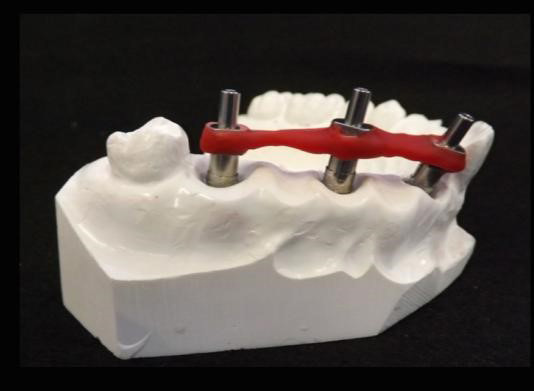
Figure 1A. Implant splint of the master mold.
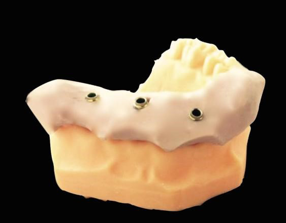
Figure 1B. Support base made of polyvinyl siloxane material adapted in the 3D printed model.
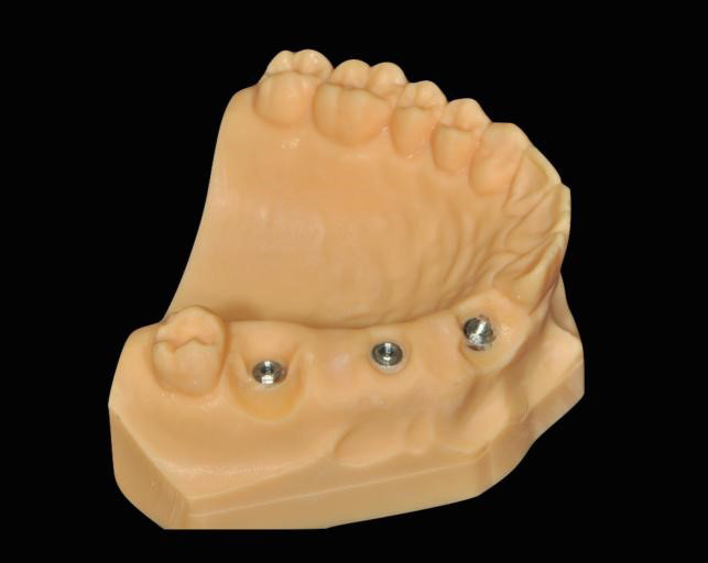
Figure 1C. 3D printed model after implant positioning.
Measurement procedure
A coordinate measuring machine (ZMC 550, Zeiss, Jena, Germany) was used to determine the distance between dental implants 22-24 (M1), 24-26 (M2) and 22-26 (M3), as shown in figure 2. The machine presents a measurement uncertainty of 4 µm, determined according to the EA- 4/02 (1999). All measurements were performed at a controlled environment with a temperature of 20±0,5oC and humidity 50±10%.
The master model was measured 5 times, and the mean values of measurements were determined as “real measurements”, whereas the conventional and 3D printed models were measured only once. The accuracy of implants position was registered as the difference between the distance values determined by the test models and the one determined by the master model (Measurement from evaluated model - Measurement from master model), named as measurement error.
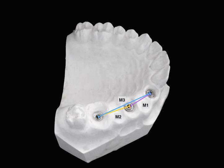
Figure 2. Distance between implants 22-24 (M1), 24-26 (M2) and 22-26 (M3) measured by a coordinate measuring machine.
Statistical analysis
Data were statistically analyzed using the software SPSS Statistics 22.0 (IBM, Armonk, USA). Kolmogorv-Smirnov and Levene tests were performed in order to evaluate the adherence to the normal curve and homogeneity of variance, respectively. As data attended to the requisites, descriptive statistics was presented as mean ± standard deviation. Statistical analysis was performed by Within ANOVA and post hoc test for pairwise comparisons with a statistical significance level at p ≤ 0.05.
Results
The position of dental implants showed a statistically higher measurement error (p≤ 0.05) for 3D printed models in comparison with conventional models, with a mean difference of 47 µm. There was significant interaction between the model type and the location of measurement, on which M2 (70±16 µm) showed lower measurement errors in comparison with M1 (136±10 µm) and M3 (155±20 µm). When analyzing data separately, M3 statistically differed between conventional and digital models, as shown in table 1.
Table 1. Mean ± standard deviation and 95% confidence interval (95% CI) (μm) of the measurement errors of conventional (CM) and digital (DM) models in M1, M2 and M3.
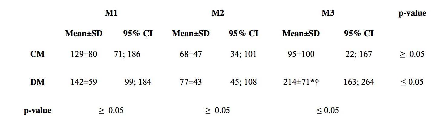
* significancia estadística en línea horizontal.
† significancia estadística en línea vertical.
Discussion
The use of a verification jig was suggested to position dental implants into a 3D printed model. It was expected that the method could be a simple and fast way to correct the implant position when any discrepancy is observed in the printed models. The main disadvantage of the technique is the need of intra-oral procedures, which require additional implant components and additional consultations6.
According to the definition of trueness and precision described in Güth et al., 201314, it was assumed that the verification jig allowed a high precision, as the low standard deviation values predicted closeness between independent measurements. However, the trueness, or closeness of agreement to the true model, was affected by the technique when the implants were separated by greater distances (M3). De La Cruz et al., 200220, showed a similar accuracy between implants positioned by verification jig and conventional impression techniques.
Nevertheless, the use of verification jig has been recommended to eliminate a misfit among dental implants when these are joined18,19. It is commonly used to record the position of implants and correct a cast procedure for both total21 and partial edentulism20. As an indexing technique, it allows to record intraorally and copy the exact relationship of implants in a master cast20.
Inaccuracies on the master cast may lead to the need of intra-oral adjustments of definitive restorations or even affect the passive fit of frameworks21. A misfit, in turn, would result in a higher susceptibility to mechanical complications, such as screw loosening and component fractures20. When the digital process is taken into consideration, different sources of errors may be attributed to the process chain17,22-25. For instance, discrepancies may be related to the lack of reference during scanning and image acquisition, as the overlap of images may occur and result in an inherent error26. Previous studies reported the influence of dental implant parameters into the scanning process27-29. Gimenez et al., 201515, reported that the accuracy of implants position in digital models was affected by the digitizing process, especially in implants separated by greater distances. Thus, the possibility of the discrepancies founded in this study being influenced by digital process cannot be discarded, as these were only found for implants separated by larger distances.
Another important variable to be considered is the use of impression materials to transfer the jig to the printed model. Lin et al., 20146, reported that the use of a verification jig is limited to cases on which adjacent analogs are correctly positioned, so that they can be used to stabilize the verification jig. To overcome this limitation, in this study the stabilization of the jig was ensured by adapting a polyvinyl siloxane support base to the adjacent teeth. It is well known, however, that impression materials may lead to the displacement of implants components and cause inaccuracy on the master cast30.
Within the limitations of this study, the use of verification jig as determined in the present study cannot be recommended. There is a need of improvement of the technique to be used to correct the implant position in 3D printed model.
Conclusion
The use of verification jig for positioning dental implants in a partial dentate 3D printed model showed greater discrepancies in comparison with conventional impressions.
Acknowledgments
The authors would like to express their gratitude to S.I.N. Implant System for the implant components donation.
Bibliografía
- Jung RE, Zembic A, Pjetursson BE, Zwahlen M, Thoma DS. Systematic review of the survival rate and the incidence of biological, technical, and aesthetic complications of single crowns on implants reported in longitudinal studies with a mean follow-up of 5 years. Clin Oral Implants Res. 2012 Oct;23(6):2-21.
- Kim JH, Kim KB, Kim WC, Rhee HS, Lee IH, Kim JH. Influence of various gypsum materials on precision of fit of CAD/CAM-fabricated zirconia copings. Dent Mater J 2015 Jan;34(1):19-24.
- de Avila ED, Barros LAB, Del'Acqua MA, Castanharo SM, Mollo FA Jr. Comparison of the accuracy for three dental impression techniques and index: an in vitro study. J Prosthodont Res 2013 Oct;57(4):268-74.
- Sorrentino R, Gherlone EF, Calesini G, Zarone F. Effect of implant angulation, connection length, and impression material on the dimensional accuracy of implant impressions: an in vitro comparative study. Clin Implant Dent Relat Res 2010 May;12(1):63-76.
- Papaspyridakos P, Hirayama H, Chen CJ, Ho CH, Chronopoulos V, Weber HP. Full-arch implant fixed prostheses: a comparative study on the effect of connection type and impression technique on accuracy of fit. Clinical Oral Implants Research 2015 Sep;27(9):1099-105.
- Lin WS, Harris BT, Metz MJ, Morton D. A technique for verifying and correcting a milled polyurethane definitive cast for nonsegmental implant restoration in an edentulous jaw. J Prosthet Dent 2014 Sep;112(3):658-62.
- Lin W-S, Harris BT, Morton D. The use of a scannable impression coping and digital impression technique to fabricate a customized anatomic abutment and zirconia restoration in the esthetic zone. J Prosthet Dent 2013 Mar;109(3):187-91.
- Lin WS, Harris BT, Zandinejad A, Morton D. Use of digital data acquisition and CAD/CAM technology for the fabrication of a fixed complete dental prosthesis on dental implants. J Prosthet Dent 2014 Jan;111(1):1-5.
- Monaco C, Evangelisti E, Scotti R, Mignani G, Zucchelli G. A fully digital approach to replicate peri-implant soft tissue contours and emergence profile in the esthetic zone. Clin Oral Implants Res 2016 Dec;27(12):1511-1514.
- Patel N. Integrating three-dimensional digital technologies for comprehensive implant dentistry. JADA 2010 Jun;141(2):20-4.
- Lee SJ, Betensky RA, Gianneschi GE, Gallucci GO. Accuracy of digital versus conventional implant impressions. Clin Oral Implants Res 2015 Jun;26(6):715-9.
- Lee CY, Wong N, Ganz SD, Mursic J, Suzuki JB. Use of an intraoral laser scanner during the prosthetic phase of implant dentistry: A Pilot Study. J Oral Implantol 2015b Aug;41(4):126-32.
- Brawek PK, Wolfart S, Endres L, Kirsten A, Reich S. The clinical accuracy of single crowns exclusively fabricated by digital workflow-the comparison of two systems. Clin Oral Investig 2013 Dec;17(9):2119-25.
- Guth JF, Keul C, Stimmelmayr M, Beuer F, Edelhoff D. Accuracy of digital models obtained by direct and indirect data capturing. Clin Oral Investig 2013 May;17(4):1201-8.
- Gimenez B, Ozcan M, Martinez-Rus F, Pradies G. Accuracy of a digital impression system based on active triangulation technology with blue light for implants: effect of clinically relevant parameters. Implant Dent 2015 Oct;24(5):498-504.
- Persson A, Andersson M, Oden A, Sandborgh-Englund G. A three-dimensional evaluation of a laser scanner and a touch-probe scanner. J Prosthet Dent 2006 Mar;95(3):194-200.
- Tapie L, Lebon N, Mawussi B, Fron-Chabouis H, Duret F, Attal JP. Understanding dental CAD/CAM for restorations-accuracy from a mechanical engineering viewpoint. Int J Comput Dent 2015;18(4):343-67.
- Knudson RC, Williams EO, Kemple KP. Implant transfer coping verification jig. J Prosthet Dent 1989 May;61(5):601-2.
- McCartney JW, Pearson R. Segmental framework matrix: master cast verification, corrected cast guide, and analog transfer template for implant-supported prostheses. J Prosthet Dent 1994 Feb;71(2):197-200.
- De La Cruz JE, Funkenbusch PD, Ercoli C, Moss ME, Graser GN, Tallents RH. Verification jig for implant-supported prostheses: A comparison of standard impressions with verification jigs made of different materials. J Prosthet Dent 2002 Sep;88(3):329-36.
- Vitale ND, Tung F, Goldstein G. A technique to verify or correct analogue position and soft tissue profile on an implant working cast. J Prosthet Dent 2009 Sep;102(3):137-40.
- Güth JF1, Keul C, Stimmelmayr M, Beuer F, Edelhoff D. Accuracy of digital models obtained by direct and indirect data capturing. Clin Oral Investig. 2013 May;17(4):1201-8.
- 23. Flügge TV1, Schlager S, Nelson K, Nahles S, Metzger MC. Precision of intraoral digital dental impressions with iTero and extraoral digitization with the iTero and a model scanner. Am J Orthod Dentofacial Orthop. 2013 Sep;144(3):471-8.
- González de Villaumbrosia P, Martínez-Rus F, García-Orejas A, Salido MP, Pradíes G. In vitro comparison of the accuracy (trueness and precision) of six extraoral dental scanners with different scanning technologies. J Prosthet Dent. 2016 Oct;116(4):543-550.
- Akyalcin, S, Dyer, D. J, English, J. D, Sar, C. Comparison of 3-dimensional dental models from different sources: Diagnostic accuracy and surface registration analysis. Am J Orthod Dentofacial Orthop. 2013 Dec;144(6):831-7.
- Rhee YK, Huh YH, Cho LR, Park CJ. Comparison of intraoral scanning and conventional impression techniques using 3-dimensional superimposition. J Adv Prosthodont 2015;7,460-7.
- Al-Abdullah K1, Zandparsa R, Finkelman M, Hirayama H. An in vitro comparison of the accuracy of implant impressions with coded healing abutments and different implant angulations.J Prosthet Dent. 2013 Aug;110(2):90-100.
- Chew AA, Esguerra RJ, Teoh KH, Wong KM, Ng SD, Tan KB. Three-Dimensional Accuracy of Digital Implant Impressions: Effects of Different Scanners and Implant Level. Int J Oral Maxillofac Implants. 2017 Jan/Feb;32(1):70-80.
- Giménez B, Özcan M, Martínez-Rus F, Pradíes G. Accuracy of a digital impression system based on parallel confocal laser technology for implants with consideration of operator experience and implant angulation and depth. Int J Oral Maxillofac Implants. 2014 Jul-Aug;29(4):853-62.
- Pujari M, Garg P, Prithviraj DR. Evaluation of accuracy of casts of multiple internal connection implant prosthesis obtained from different impression materials and techniques: an in vitro study. J Oral Implantol 2014;40,137-45.

Reconocimiento-NoComercial-CompartirIgual
CC BY-NC-SA
Esta licencia permite a otros entremezclar, ajustar y construir a partir de su obra con fines no comerciales, siempre y cuando le reconozcan la autorÍa y sus nuevas creaciones estÉn bajo una licencia con los mismos tÉrminos.