RESUMEN
Introducción: La microabrasión se describe como un procedimiento realizado sobre el esmalte dental en el cual mediante la utilización de un agente ácido y un agente abrasivo se logra corregir alteraciones cromáticas superficiales. Algunos estudios demuestran como los parámetros de tiempo, número de aplicaciones y la presión ejercida, influyen en la cantidad de esmalte eliminado. Objetivo: Establecer el espesor de esmalte dental eliminado según la capacidad abrasiva de 9 tratamientos químico mecánicos, mediante estereomicroscopia. Materiales y métodos: Con el aval del comité de ética de la Facultad de Odontología de la Universidad Nacional de Colombia, se recolectaron 90 terceros molares bajo consentimiento informado y se mantuvieron almacenados bajo los parámetros de la norma ISO 11405. Sobre bloques de acrílico se fijaron las mitades linguales de las coronas dentales, creando sobre ellas superficies planas mediante serie de lijas con irrigación y tomando impresiones con silicona de adición. Se distribuyeron de forma aleatoria en 9 grupos (n 10). Cada grupo recibió un tratamiento por un periodo de 30 segundos: G1: Opalustre® (Ultradent), G2: Piedra pómez y ácido fosfórico al 37% (Ultra-Etch®, Ultradent), G3: Piedra pómez, glicerina y ácido fosfórico al 37% (Ultra-Etch®, Ultradent), G4: Fresas de halo amarillo (Komet), G5: Fresas de halo blanco (Komet), G6: Discos Sof-Lex® (3M), color amarillo, G7: Discos Sof-Lex® (3M), color amarillo y amarillo claro, G8: Arenado, y G9: Puntas ultrasónicas Perfect Margin (Acteon). El espesor de desgaste creado fue medido utilizando un estéreo microscopio con un aumento de 10X. Los datos recolectados se analizaron a través de las pruebas de Kruskal-Wallis (p≤0.05) para comparar todos los grupos y la prueba U de Mann-Whitney (p≤0.05) para comparaciones individuales. Resultados: Independientemente del tratamiento realizado todos los grupos presentaron un desgaste del esmalte. El mayor desgaste se registró para el grupo tratado con fresa de halo amarillo (122,66 ± 22,64µm) y el menor desgaste para el grupo de arenado (11,5 ± 2,36µm). Se presentó diferencia estadísticamente significativa entre todos los grupos. Conclusiones: Bajo las limitaciones del presente estudio se puede concluir: La mayor microabrasión en esmalte se produjo con fresas de grano extrafino (halo amarillo) y el menor desgaste se produjo con arenado.
Palabras clave: Esmalte dental; pulido dental / métodos; esmalte dental / efectos farmacológicos; microabrasión / métodos de esmalte; abrasión de aire.
ABSTRACT
Introduction: Microabrasion is described as a procedure performed on tooth enamel in which the use of an acidic agent and an abrasive agent can correct surface chromatic alterations. Some studies show how the parameters of time, number of applications and the pressure exerted influence the amount of enamel removed. Objective: To establish the thickness of tooth enamel removed according to the abrasive capacity of 9 mechanical chemical treatments, using stereomicroscopy. Materials and methods: With the endorsement of the ethics committee of the School of Dentistry of the National University of Colombia, 90 third molars were collected under informed consent and kept stored under the parameters of ISO 11405. Acrylic blocks were fixed the lingual halves of the dental crowns, creating on them flat surfaces by means of series of sandpaper with irrigation and taking impressions with silicone of addition.They were distributed randomly in 9 groups (n 10). Each group was treated for a period of 30 seconds: G1: Opalustre® (Ultradent), G2: Pumice and 37% phosphoric acid (Ultra-Etch®, Ultradent), G3: Pumice, glycerin and phosphoric acid 37 % (Ultra-Etch®, Ultradent), G4: Yellow halo strawberries (Komet), G5: White halo strawberries (Komet), G6: Sof-Lex® discs (3M), yellow color, G7: Sof-Lex discs ® (3M), yellow and light yellow, G8: Sandblasted, and G9: Perfect Margin ultrasonic tips (Acteon). The wear thickness created was measured using a stereo microscope with an increase of 10X. The collected data were analyzed through the Kruskal-Wallis tests (p≤0.05) to compare all groups and the Mann-Whitney U test (p≤0.05) for individual comparisons. Results: Regardless of the treatment performed, all groups presented enamel wear. The highest wear was recorded for the group treated with yellow halo strawberry (122.66 ± 22.64µm) and the lowest wear for the sandblasting group (11.5 ± 2.36µm). There was a statistically significant difference between all groups. Conclusions: Under the limitations of the present study, it can be concluded: The greatest microabrasion in enamel was produced with strawberries of extra-fine grain (yellow halo) and the least wear occurred with sandblasting.
Keywords: Dental enamel; dental polishing / methods; dental enamel / drug effects; enamel microabrasion / methods; air abrasion.
RESUMO
Introdução: A microabrasão do esmalte dental é descrita como um procedimento realizado no esmalte dentário, no qual o uso de um agente ácido e um abrasivo pode corrigir alterações cromáticas na superfície. Alguns estudos mostram como os parâmetros de tempo, número de aplicações e pressão exercida influenciam na quantidade do esmalte removido. Objetivo: Estabelecer a espessura do esmalte dentário removido de acordo com a capacidade abrasiva de 9 tratamentos químicos ou mecânicos, utilizando estereomicroscopia. Materiais e métodos: Com o aval do comitê de ética da Faculdade de Odontologia da Universidade Nacional da Colômbia, 90 terceiros molares hígidos foram coletados sob consentimento informado e mantidos armazenados sob os parâmetros da norma ISO 11405. Em blocos de acrílico foram fixadas as metades linguais das coroas dentárias, criando sobre elas superfícies planas por meio de séries de lixa mais irrigação e toma de impressões com silicone de adição. Eles foram distribuídos aleatoriamente em 9 grupos (n= 10). Cada grupo foi tratado por um período de 30 segundos: G1: Opalustre® (Ultradent), G2: Pedra-pomes e ácido fosfórico a 37% (Ultra-Etch®, Ultradent), G3: Pedra-pomes, glicerina e ácido fosfórico 37 % (Ultra-Etch®, Ultradent), G4: brocas diamantadas halo amarelo (Komet), G5: brocas diamantadas halo branco (Komet), G6: discos Sof-Lex® (3M), cor amarelo, G7: discos Sof-Lex ® (3M), amarelo e amarelo claro, G8: jateamento e G9: pontas ultra-sônicas Perfect Margin® (Acteon). A espessura de desgaste criada foi medida usando um microscópio estéreo com um aumento de 10X. Os dados coletados foram analisados pelos testes de Kruskal-Wallis (p≤0,05) para comparar todos os grupos e pelo teste U de Mann-Whitney (p≤0,05) para comparações individuais. Resultados: Independentemente do tratamento realizado, todos os grupos apresentaram desgaste do esmalte. O maior desgaste foi registrado para o grupo tratado com broca diamantada com halo amarelo (122,66 ± 22,64 µm) e o menor desgaste para o grupo de jateamento (11,5 ± 2,36 µm). Houve diferença estatisticamente significante entre todos os grupos. Conclusões: Sob as limitações do presente estudo, pode-se concluir que a maior microabrasão no esmalte foi produzida com brocas de grão extra-fino (halo amarelo) e o menor desgaste ocorreu com o jateamento.
Palavras-chave: Esmalte dentário; polimento dentário / métodos; esmalte dentário / efeitos de drogas; microabrasão de esmalte / métodos; abrasão a ar.
INTRODUCTION
Microabrasion is a technique to eliminate chromatic and / or structural surface alterations in the enamel. The first related report was made by Walter Kane in 1916, who proposed the use of hydrochloric acid (HCl) with and without heat to remove brown spots caused by fluorosis. For the treatment of the same type of spots, McCloskey (1984) used 18% HCl with a subsequent polishing with pumice stone and a waiting time to achieve remineralization of the surface by the saliva1,2. Subsequently, Croll and Cavanaugh (1986), describe a technique for the treatment of any type of alteration in dental color, which included the joint use of 18% HCl with pumice stone, then a prophylaxis with fluoridated paste and finally a polishing with an ultra-thin disk of aluminum oxide3.
Mondelli et al., (1995), describe an alternative for the microabrasion technique using phosphoric acid (H3PO4) in 37% gel in combination with extra fine pumice stone in a 1: 1 ratio4, this technique has certain advantages over those already described, because for the dentist the H3PO4 has a low cost, easy acquisition and handling5. In addition, it is available in the office because it is used in different techniques such as the preparation of the surface for restoration with materials that require adhesion6 and in the dental preparation for bonding the resin to the brackets. Additionally, H3PO4 is less aggressive with soft tissues than HCl, in case of accidental contact4.
Some studies have established that the factors quantity of applications, time of use, and pressure exerted, have a direct impact on the amount of enamel removed5,7. However, not all parameters are included in the studies, so differences in the results obtained are found in the literature. For example, with the use of phosphoric acid, wear of 5.7 µm (+/- 1.8)8 and with 18% HCl wear of 100 µm (+/- 47 µm) have been reported (8) ; phosphoric acid followed by pumice, wear of 142.87 microns (5), while in the mixture of HCl with pumice between 295.5 microns (5) and up to 360 µm (+/- 130)8.
Another technique used to realize microabrasion, which is described in the literature, is the use of fine grain strawberries between 60 and 80 µm. In the work done by Agudelo and Calvo9, the microabrasion was measured on enamel without alterations, performing 3 sessions for 10 seconds (5 s with yellow strawberry and 5 s with white strawberry) and exerting a pressure between 20 or 30 g, establishing wear between 200 and 400 µm.
The same happens with sandblasting techniques, created as alternatives to the use of phosphoric acid in the enamel conditioning. It uses an air flow of 30 psi and particles of aluminum oxide (50-90 µm) that create roughness for the micromechanical retention of the resins, but the binding values are lower than those of acid etching. Pressure, time and their interaction have significant effects10.
Many treatments routed at conditioning or eliminating surface defects are empirically done without control of the probable variables, being able to achieve severe wear, which exceed the limits of microabrasion. Currently, technological progress allows us to observe and quantify the changes in the tissue and from these verify through a risk-benefit model, how relevant is the performance of each treatment and how these could be improved, adjusting the necessary parameters to reduce the amount of Enamel removed in each process.
The objective of the present study was to establish the thickness of tooth enamel removed using a stereo microscope, according to the abrasive capacity of nine chemical-mechanical treatments.
Materials and methods
After the approval of the ethics committee of the Faculty of Dentistry of the National University of Colombia, a sample of 90 molar third parties was obtained, obtained with informed consent from systemically healthy patients and whose exodontics were performed by orthodontic or periodontal indications. Those with caries, fillings or alterations in enamel formation were excluded. The collected teeth were washed with water and the soft tissue remains were removed.
The teeth were stored according to ISO 11405, in containers with a 0.5% Chloramine T solution at 4°C for 8 days. Subsequently, the chloramine was exchanged for distilled water with refills every week until the time of the assembly of the test. For the experiment, coronal cuts were made meso-distally to obtain the lingual halves that were fixed on acrylic cubes attached to threaded attachments. (figure 1. A). These test bodies were left in the Hygrobath at 37°C (+/- 2) with 70% relative humidity.
In order to achieve flat test areas in the enamel of the lingual surfaces, series of low speed and low cooling sandpaper were used, ending with fine sandpaper of the OptiDisc® (Kerr) system. Then, cylinders 21 mm in diameter and 21 mm high in acetate were manufactured, which were used as cuvettes for printing in polyvinyl siloxane, which served as a reference for wear measurements. Then a cut with scalpel blade No.11 was made in the printout, which coincided with the distal half meso of the test area and thus allowed measurements with stereo microscope (figure 1. B).
The test areas achieved in the enamel, were divided into two mesial and distal halves, drawing a line with graphite pencil. The mesial halves were used as controls and were covered with adhesive tape and the distal halves served as an application area for the different microabrasion treatments (figure 1. C).
The teeth were randomly divided into 9 groups (n= 10). All groups received the specific treatment by the same operator, for a time of 30 seconds and a pressure of 120 to 150g controlled by the use of a pressure standardizer.
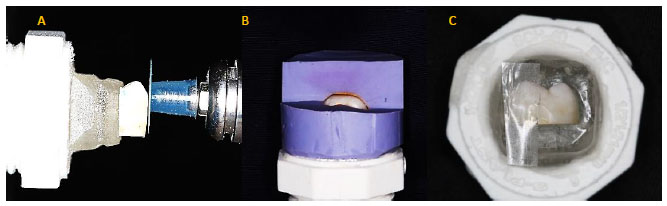
Figure 1. Preparation of sample bodies. A, Execution of planimetry area on the surface of dental enamel with sandpaper and OptiDisc® system (Kerr). B, Silicone impression of adding one of the experimental teeth, where you can see the window created for the measurement. C, Experiment tooth, where the control surface covered by adhesive tape is observed.
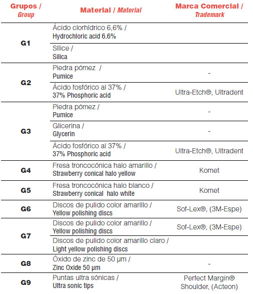
The experimental groups were: G1. Microabrasion with pastes based on hydrochloric acid (Opalustre®, Ultradent), G2. Microabrasion with pumice stone and 37% phosphoric acid (Ultra-Etch®, Ultradent), G3. Microabrasion with pumice stone, glycerin and 37% phosphoric acid (Ultra-Etch®, Ultradent), G4. Preparation with strawberries of yellow halo (Komet), G5. Preparation with white halo strawberries (Komet), G6. Preparation with yellow Sof-Lex® (3M) discs, G7. Combination of Sof-Lex® (3M) discs, yellow and light yellow, G8. Sandblasting and G9. Perfect Margin® (Acteon) ultrasonic system tips. Table 1 presents the main information about the products used in this study.
Table 1.Distribution of groups and materials used.
In group 1, microabrasion paste based on hydrochloric acid (Opalustre®, Ultradent) was placed, leaving the test area for 10 seconds; then 2 applications of 10 seconds with rubber cup and low speed were made, executing circular movements at 15,000 rpm and a pressure between 120-150 g (figure 2. A). Then it was washed with triple syringe spray.
Group 2 received the 37% phosphoric acid mixture (Ultra-Etch®, Ultradent), with the pumice stone and rubbed for 30 seconds, then washed with triple syringe spray.
In group 3, 37% phosphoric acid (Ultra-Etch®, Ultradent) was applied, with pumice in glycerin and rubbed for 30 seconds, then washed with triple syringe spray.
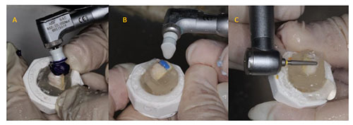
Figure 2. Realization treatments on the dental surface. A, Opalustre® treatment. B, Treatment with pumice and phosphoric acid (Ultra-Etch®). C, Treatment with yellow truncated cone strawberry.
In group 4, fine yellow-cone strawberry trunks (extra-fine grain, Komet) were used, maintaining a pressure between 20 and 30g for 3 sessions of 10 seconds at 15,000 rpm.
In group 5, white halo truncated cone strawberries (ultra-fine grain, Komet) were used under the same conditions as group 4.
In group 6, yellow Sof-Lex® (3M) disc was used with irrigation under the same conditions of groups 4 and 5.
In group 7 consecutively used Sof-Lex® (3M) discs, first yellow for 15 seconds and then light yellow for 15 seconds for a total of 30 seconds. The disks slid unidirectionally.
Group 8 received the 50 micron aluminum oxide sandblasting technique by pressing for 30 seconds at a distance of 5 mm. Finally it was washed profusely with water.
Group 9 received the Sonic System tip technique with 70 lb. air pressure and Perfect Margin® (Acteon) cylindrical tip, for 30 seconds unidirectionally.
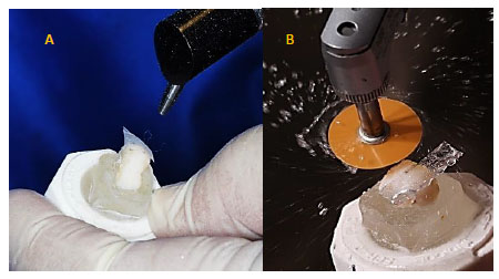
Figure 3. Treatments performed on dental surfaces. A, Sandblasting treatment (50micron aluminum oxide). B, Treatment with yellow SofLex discs.
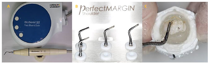
Figure 4. Ultrasonic tip system used. A, BioSonic S1 ultrasonic scaler system, Coltene. B, PerfectMargin Shoulder ultrasonic tips. C, Treatment with ultrasonic tips.
Ending each treatment on the respective teeth, the silicone matrix was repositioned, 15 minutes were expected to achieve elastic recovery of the material, before observation with 10X Opticks stereo microscope. Digital images were taken of the gap corresponding to enamel wear with each treatment, making two measurements using the Motic 3.2 image analyzer. These data were collected for subsequent statistical analysis.
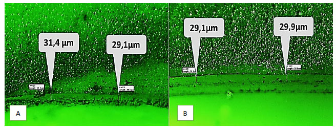
Figure 5. 80X stereomicroscopy images, analyzed with Motic 3.2. A, Aspect of the gap corresponding to the abrasion achieved with Opalustre® in 30 seconds. B, Gap corresponding to the microabrasion obtained with pumice stone, glycerin and phosphoric acid.
Once the study was finished, the specimens were taken to calcination.
The data analysis was carried out through the non-parametric statistical tests of Kruskal Wallis (p≤0.05) which was used to compare if there were significant differences between all groups and the Mann-Whitney U test (p≤0,05) to make comparisons between pairs.
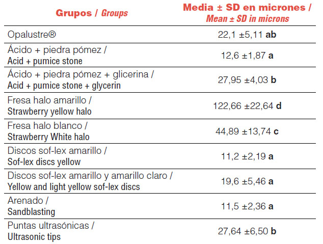
Table 2. Mean and standard deviation of enamel wear (µm) for each treatment performed, groups with different letters presented statistical differences (p≤0.05).
Graphic 1. Values of mean wear generated by each treatment performed for 30 seconds.
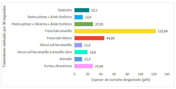
Chart 2 shows that all the treatments used to perform microabrasion generated wear. The highest wear was recorded for the group treated with yellow halo strawberry (122.66 ± 22.64 µm) and the lowest wear for the group treated with sandblasting (11.5 ± 2.36 µm).
It was presented a statistically significant difference when making a comparison between all the experimental groups (p <0.01). At the even comparisons between the groups there were no significant differences of some such as Opalustre® versus Sof-Lex® yellow + light yellow discs (p = 0.289), as well as between the pumice stone + glycerin + phosphoric acid group compared to the group of Ultrasound.
Discussion
The traditional microabrasion technique uses a combination of a hydrochloric acid agent and an abrasive element. It has been described as a non-restorative, conservative and safe technique11, its main objective is the removal of color changes, pigmentations or alterations in the dental surface with a minimum enamel loss of up to 250 µm5. Other forms of microabrasion described have been the use of fine and ultrafine strawberries, sandblasting, sonic tips, among others9,12.
In the present work, when observing under stereomicroscopy, different levels of enamel wear were found in the groups, being significantly higher in the group treated with fine strawberries (yellow halo) and ultrafine (white halo), in a total time of 30 seconds and with a gentle pressure, which allows to establish that under similar conditions, this method could be used for microabrasion for a time not exceeding one minute and should be supplemented with Sof-Lex® discs.
Opalustre® is a 6.6% hydrochloric acid viscous paste that contains silica microparticles in a water-soluble base. It is one of the most commonly used commercial substances for microabrasion13-16. Sundfeld et al., Showed that in extracted teeth treated with Opalustre®, making 1 to 10 applications for 1 minute on each tooth, wear from 25 to 200 µm14 could be generated, while Rodrigues et al. Found that for a 60 second application generated wear of 26.96 ± 5.70 µm14. In the present work, by applying Opalustre® with a rubber cup, at a pressure of 150g for a period of 30 seconds, wear was achieved between 22.1 ± 5.11 µm, it was estimated that under similar conditions a Maximum of 10 applications.
With the mixture of phosphoric acid and pumice stone, an immediate reaction was observed, with different values of enamel wear being reported in the literature, due to methodological variability. In the work carried out by Mendes et al., The wear generated was 142.87 µm, after 10 applications of 5 seconds each, for a total of 50 seconds17. Rodrigues et al., Found that for a 60-second application the wear generated was 27.65 ± 6.57 µm14. This result is similar to that present in the group that included glycerin as a vehicle for the mixture of phosphoric acid and pumice, with an average wear of 27.95 ± 4.03 µm (which in turn is close to those reported for Opalustre®).
When comparing the phosphoric acid +pumice+glycerin(27.95±4.03µm) groups versus the phosphoric acid + pumice (12.6±1.87 µm) group, it can be thought that the glycerin group avoids that the acid is neutralized generating greater wear. However, no similar studies were found that match these results, so it is recommended to perform new work with this mixture that could be a low cost and lower risk alternative, compared to hydrochloric acid based pastes.
The use of stain removal techniques with fine grain strawberries is described as an accompaniment to microabrasion and they are used when the stains present are at a greater depth18. In the present investigation these are the groups with the highest levels of wear generated, 44.89± 13.74 µm for white halo strawberries, and 122.66 ± 22.64 µm for yellow halo strawberry.
The use of polishing discs, Sof-Lex®, yellow and yellow + light yellow, allowed to show a wear of 19.6 µm, this should be taken into account, because this type of treatment is usually used at the end of any microabrasion technique with the aim of refining the enamel, generating complementary wear.
Wear with the use of abrasive air (sandblasting) can vary the results depending on the particle size used, the air pressure, the diameter of the nozzle and the distance between the nozzle and the dental surface12. In the study conducted by Lambrechts et al., It is mentioned that treatment with abrasive air on the vestibular dental surface can generate wear of up to 595 µm at the deepest points12. However, it does not mention the conditions under which the treatment is performed (time, air pressure, etc.). In the present investigation using 50 µm aluminum oxide as a particle at a distance of 5mm, with an air pressure of 35 pounds for 30 seconds, an average wear of 15.3 µm was obtained.
The ultrasonic tip system has not been very studied in terms of microabrasion, however, the results of this study, show it as another option generating an average wear of 27.64 ± 6.50 µm, for 30 seconds.
Having as reference the recommended range for the microabrasion technique (250-300µm) and the conditions of the present work, different protocols can be suggested, for example, abrasion with fine grain strawberries, it can take up to 2 sessions of 30 seconds, for ultra-fine strawberries up to 6 sessions of 30 seconds, for pastes based on hydrochloric acid and the mixture of phosphoric acid with pumice and glycerin used in this study, up to 10 applications of 30 seconds and for the other methods up to 20 applications of 30 seconds.
Conclusions
With the limitations of the present study in mind, it can be concluded that: The enamel wear range was between 11 and 122 microns in 30-second applications. The group with the highest microabrasion in enamel was that of fine strawberries and the group with the lowest microabrasion was sandblasting.
Interests conflict
The authors declare no conflict of interest in the presentation of data, preparation and publication of this article.
Bibliografía
- McCloskey, RJ. A technique for removal of fluorosis stains. The Journal of the American Dental Association. 1984. 109(1), 63–64.
- Álvarez M, Quiroz K, Rodriguez V, Castelo RM. Dental microabrasion in children: An esthetic alternative. Odontol. Sanmarquina. 2009; 12(2): 86-89
- Croll TP, Cavanaugh R. Enamel color modification by controlled hydrochloric acid and pumice abrasion. Quintessence Int. 1986; 7 (2): 26-28.
- Mondelli J, Mondelli RFL, Bastos MT, Franco EB. Microabrasão com ácido fosfórico. Rev. bras. de Odont.1995. 52(3): 20-22.
- Meireles SS, Andre Dde A, Leida FL, Bocangel JS, Demarco FF. Surface roughness and enamel loss with two microabrasion techniques. J Contemp Dent Pract. 2009;10:58–65.
- Bertacci A, Lucchese A, Taddei P, Gherlone EF, Chersoni S. Enamel structural changes induced by hydrochoric and phosphoric acid treatment. J Appl Biomater Funct Mater. 2014;12(3):240 -247.
- Ardu S, Benbachir, Sttavridakis M, Dietshi D, Krejci, Feilzer. A combined chemo-mechanical approach for aestetic managemof superficial enamel defects. A British Dental Journal. 2009;206(4): 205-208.
- Tong LSM, Pang MKM, Mok NYC, King NM, Wei SHY. The effects of etching, micro-abrasion, and bleaching of surface enamel. Journal of Dental Restauration.1993;72(1):67-71.
- Agudelo LJ. Efecto de dos sistemas de microabrasión en el espesor del esmalte dental. [Tesis]. Bogotá: Universidad Nacional de Colombia. 2017.
- van Waveren Hogervorst WL, Feilzer AJ, Prahl-Andersen B. The air-abrasion technique versus the conventional acid-etching technique: A quantification of surface enamel loss and a comparison of shear bond strength. American Journal of Orthodontics and Dentofacial Orthopedics. 2000;117(1):20-6.
- Pini NIP, Costa R, Bertoldo CE, Aguiar FH, Lovadino JR, D Alves. Enamel morphology after microabrasion with experimental compounds. Contemp Clin Dent. 2015;6(2):170–175.
- Lambrechts P, Mattar D, De Muck J, Bergmans L, Peumans M, Vanherle G, Van Merrbeeck B. Air-abrasion enamel microsurgery to treat enamel White spot lesions of traumatic origin. Masters of esthetic dentistry. 2002. 14 (3) 167-187.
- Sundfeld RH, Briso ALF, Mauro SJ. Smile recovery. IV. External whitening of traumatized teeth. J Bras Clin Estet Odontol 2000;5:29-35.
- Rodrigues MC, Mondelli RFL, Oliveira GU, Franco EB, Baseggio W, Wang L. Minimal alterations on the enamel surface by micro-abrasion: in vitro roughness and wear assessments. J. Appl. Oral Sci. [Internet]. 2013. [cited 2018 Oct 20] ; 21 (2): 112-117.
- Paic M, Sener B, Schug J, Schmidlin PR. Effects of microabrasion on substance loss, surface roughness, and colorimetric changes on enamel in vitro. Quintessence International. 39 (6): 517-522.
- Bertoldo C, Lima D, Fragoso L, Ambrosano G, Aguiar F, Lovadino J. Evaluation of the effect of different methods of microabrasion and polishing on surface roughness of dental enamel. Indian J Dent Res. 2014 May-Jun;25(3):290-3
- Mendes RF, Mondelli J, Freitas CA. Avaliação da quantidade de desgaste do esmalte dentário submetido à microabrasão. Rev Facul Odont Bauru. 1999;7:6
- Sundfeld RH, Croll TP, Fraga AL, Sversut De Alexandre R, Neto DS. Considerations about enamel microabrasion after 18 years. American Journal of Dentistry. 2007. 20:67-72

Reconocimiento-NoComercial-CompartirIgual
CC BY-NC-SA
Esta licencia permite a otros entremezclar, ajustar y construir a partir de su obra con fines no comerciales, siempre y cuando le reconozcan la autorÍa y sus nuevas creaciones estÉn bajo una licencia con los mismos tÉrminos.