Expresión génica de BMPR1, BMPR2, y TGFBR1 en dentina humana tratados con diferentes soluciones desmineralizantes y adhesión de las células de papila apical cultivada
DOI:
https://doi.org/10.29166/odontologia.vol23.n2.2021-e3440Palabras clave:
Diferenciación Celular, Adhesión Celular, EGTA, Endodoncia Regenerativa, Células MadreResumen
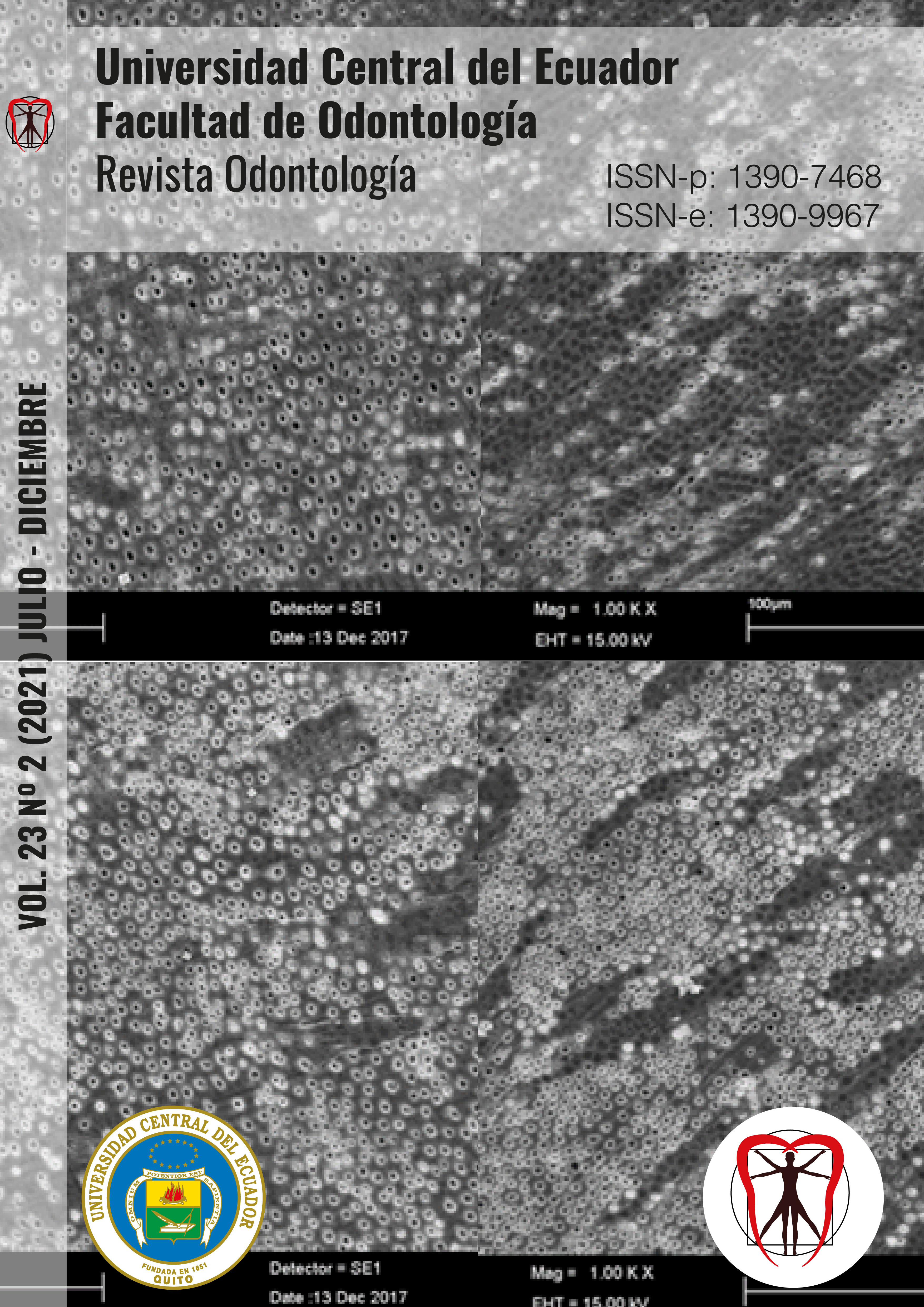
El éxito de los procedimientos de endodoncia regenerativa depende de la descontaminación del espacio endodóntico, la presencia de células mesenquimales indiferenciadas, la liberación de factores de crecimiento que actúan como moléculas señalizadoras para la atracción, proliferación y diferenciación celular; y de la presencia de un “scaffold”, que brinde soporte a la organización y vascularización del tejido recién formado. Objetivo. Analizar soluciones quelantes específicas de calcio EGTA, Ácido Cítrico y EDTA en la liberación de factores de crecimiento (BMPR1, BMPR2 y TGFBR1) presentes en la dentina como la adhesión célular. Materiales y Métodos. Se obtuvieron discos de dentina de premolares y terceros molares humanos, después de la imnersión por 1 minuto con EDTA, EGTA y Ácido Cítrico, se sembraron las células. La adhesión celular se evaluó a las 48 horas usando MEV. La expresión génica se determinó a los 7 y 21 días de cultivo celular. El análisis estadístico fue ANOVA seguido de Tukey, con un nivel de significancia del 5%. Resultados. A los 21 días, el tratamiento con EGTA aumento significativamente BMPR1 en comparación con PBS. Ninguna condición experimental alteró la expresión de BMPR2. La expresión de TGBR1 a los 21 días aumentó significativamente al tratamiento con EGTA. Aunque no hubo una distribución uniforme de células a lo largo de los discos, se observó adhesión celular en todos los grupos experimentales. Conclusiones. El tipo de solución desmineralizante interfiere con la cantidad liberada de factores de crecimiento BMPR1 y TGFBR1, de la dentina humana, y no interfiere en la adhesión celular.
Descargas
Citas
Bose R, Nummikoski P, Hargreaves K. A retrospective evaluation of radiographic outcomes in immature teeth with necrotic root canal systems treated with regenerative endodontic procedures. J Endod 2009;35:1343-9.
Rafter M. Apexification: a review. Dent Traumatol 2005;21:1–8.
Witherspoon D, Small J, Regan J, et al. Retrospective Analysis of Open Apex Teeth Obturated with Mineral Trioxide Aggregate. J Endod. 2008:34:1171-6.
Shah N, Logani A, Bhaskar U, Aggarwal V. Efficacy of revascularization to induce apexification/apexogensis in infected, nonvital, immature teeth: a pilot clinical study. J Endod. 2008 Aug;34(8):919-25.
Wang X, Thibodeau B, Trope M, Lin LM, Huang GT. Histologic characterization of regenerated tissues in canal space after the revitalization/revascularization procedure of immature dog teeth with apical periodontitis. J Endod. 2010 Jan;36(1):56-63.
Bansal R, Bansal R. Regenerative endodontics: a state of the art. Indian J Dent Res. 2011 Jan-Feb;22(1):122-31.
Zhang W, Yelick PC. Vital pulp therapy-current progress of dental pulp regeneration and revascularization. Int J Dent. 2010;2010:856087.
Huang GT, Sonoyama W, Liu Y, Lin H, Wang S, Shi S. The hidden treasure in apical papilla: the potential role in pulp/ dentin regeneration and bioroot engineering. J Endod. 2008 Jun;34(6):645-51.
Gomes-Filho JE, Duarte PC, Ervolino E, Mogami Bomfim SR, Xavier Abimussi CJ, Mota da Silva Santos L, et al. Histologic characterization of engineered tissues in the canal space of closed- apex teeth with apical periodontitis. J Endod. 2013.
AAE- American Association of Endodontics. Considerations for Regenerative Procedures [citado 1 out. 2013]. Disponível em: http://www.aae.org/clinicalresources/regenerativeendodontics/considerations-for- regenerativeprocedures.aspx.
Wigler R, Kaufman AY, Lin S, et al. Revascularization: a treatment for permanent teeth with necrotic pulp and incomplete root development. J Endod 2013;39:319–26.
Clarkson RM, Moule AJ. Sodium hypochlorite and its use as an endodontic irrigant. Aust Dent J. 1998 Aug;43(4):250-6.
Gavini G, Siqueira EL, Lemos EM, Amaral KF. Substancias Químicas. In: MACHADO MEL. Endodoncia – Ciencia y Tecnologia. Caracas: AMOLCA; 2016. p. 539-577.
Trevino EG, Patwardhan AN, Henry MA, et al. Effect of irrigants on the survival of human stem cells of the apical papilla in a platelet-rich plasma scaffold in human root tips. J Endod 2011;37:1109–15.
Tziafas D, Alvanou A, Panagiotakopoulos N, Smith AJ, Lesot H, Komnenou A, Ruch JV. Induction of odontoblast-like cell differentiation in dog dental pulps after in vivo implantation of dentine matrix components. Arch Oral Biol. 1995 Oct;40(10):883-93.
Zhao S, Sloan AJ, Murray PE, Lumley PJ, Smith AJ. Ultrastructural localisation of TGF-beta exposure in dentine by chemical treatment. Histochem J. 2000 Aug;32(8):489-94.
Pang N, Seung Jong Lee, Euiseong Kim, et al. Effect of EDTA on Attachment and Differentiation of Dentária Pulp Stem Cells. J Endod. 2014 Jun;40:5.
Galler k, Buchalla W, Hiller K, et al. Influence of Root Canal Disinfectants on Growth Factor Release from Dentin. J Endod. 2015:41:363-8.
Santibañez JF, Quintanilla M, Bernabeu C. TGF-β/TGF-β receptor system and its role in physiological and pathological conditions. Clin Sci (Lond). 2011 Sep;121(6):233-51.
Cassidy N, Fahey M, Prime SS, Smith AJ. Comparative analysis of transforming growth factor-beta isoforms 1-3 in human and rabbit dentine matrices. Arch Oral Biol. 1997 Mar;42(3):219-23.
Toyono T, Nakashima M, Kuhara S, Akamine A. Expression of TGF-beta superfamily receptors in dental pulp. J Dent Res. 1997 Sep;76(9):1555-60
Sloan AJ, Smith AJ. Stimulation of the dentine-pulp complex of rat incisor teeth by transforming growth factor-beta isoforms 1-3 in vitro. Arch Oral Biol.1999 Feb;44(2):149-56.
Shirakawa M, Shiba H, Nakanishi K, Ogawa T, Okamoto H, Nakashima K, Noshiro M, Kato Y. Transforming growth factor-beta-1 reduces alkaline phosphatase mRNA and activity and stimulates cell proliferation in cultures of human pulp cells. J Dent Res. 1994 Sep;73(9):1509-14.
Shiba H, Fujita T, Doi N, Nakamura S, Nakanishi K, Takemoto T, Hino T, Noshiro M, Kawamoto T, Kurihara H, Kato Y. Differential effects of various growth factors and cytokines on the syntheses of DNA, type I collagen, laminin, fibronectin, osteonectin/secreted protein, acidic and rich in cysteine (SPARC), and alkaline phosphatase by human pulp cells in culture. J Cell Physiol. 1998 Feb;174(2):194-205.
Melin M, Joffre-Romeas A, Farges JC, Couble ML, Magloire H, Bleicher F. Effects of TGFbeta1 on dental pulp cells in cultured human tooth slices. J Dent Res. 2000 Sep;79(9):1689-96.
Kim M, Choe S. BMPs and their clinical potentials. BMB Rep. 2011 Oct;44(10):619-34.
Solofomalala GD, Guery M, Lesiourd A, Le Huec JC, Chauveaux D, LaVenetre O. Bone morphogenetic proteins: from their discoveries till their clinical applications. Eur J Orthop Surg Traumatol. 2007;17(6):609-15.
Chen D, Zhao M, Mundy GR. Bone morphogenetic proteins. Growth Factors. 2004 Dec;22(4):233-41. Review. PubMed PMID: 15621726.
Matthews SJ. Biological activity of bone morphogenetic proteins (BMP’s). Injury. 2005 Nov;36 Suppl 3:S34-7.
Ivica A, Deari S, Patcas R, Weber FE, Zehnder M. Transforming Growth Factor Beta 1 Distribución y Contenido en la Dentina raíz de jóvenes maduros e inmaduros premolares humanos. J Endod. 2020 mayo;46(5):641-647.
Galler KM, Widbiller M, Buchalla W, Eidt A, Hiller KA, Hoffer PC, Schmalz G. EDTA conditioning of dentine promotes adhesion, migration and differentiation of dental pulp stem cells. Int Endod J. 2016 Jun;49(6):581-90.
Hashimoto K, Kawashima N, Ichinose S, Nara K, Noda S, Okiji T. EDTA Treatment for Sodium Hypochlorite-treated Dentin Recovers Disturbed Attachment and Induces Differentiation of Mouse Dental Papilla Cells. J Endod. 2018 Feb;44(2):256-262.
Serper A, Çalt S. The demineralizing effects of EDTA at different concentrations and pH. J Endod 2002;28:501-2.
Sousa-Neto M, Passarinho-neto J, Carvalho-júnior J, et al. Evaluation of the Effect of EDTA, EGTA and CDTA on Dentin Adhesiveness and Microleakage with Different Root Canal Sealers. Braz Dent J (2002) 13(2): 123-128.
Sousa S e Silva T. Desmineralization effect of EDTA, EGTA, CDTA and citric acido n root dentin: a comparative study. Braz Oral Res 2005 19:3.
Calt S, Serper A. Smear layer removal by EGTA. J Endod. 2000 Aug;26(8):459-61.
Scelza MF, Pinheiro Daniel RLD, Santos EM, Jaeger MMM. Cytotoxic effects of 10% citric acid and EDTA-T used as root canal irrigants: an in vitro analysis. J Endod 2001;27:741-3.
Scelza MFZ, Teixeira AM, Scelza P. Decalcifying effect of EDTA-T, 10% citric acid and 17% EDTA on root canal dentin. Oral Surg Oral Med Oral Pathol Oral Radiol 2003;95:234-6.
Amaral KF, Rogero MM, Fock RA, Borelli P, Gavini G. Cytotoxicity analysis of EDTA and citric acid applied on murine resident macrophages culture. Int Endod J.2007 May;40(5):338-43.
Smith AJ, Leaver AG. Non-collagenous components of the organic matrix of rabbit incisor dentine. Arch Oral Biol. 1979;24(6):449-54.
Mazzoni A, Pashley DH, Tay FR, Gobbi P, Orsini G, Ruggeri A Jr, Carrilho M, Tjäderhane L, Di Lenarda R, Breschi L. Immunohistochemical identification of MMP-2 and MMP-9 in human dentin: correlative FEI-SEM/TEM analysis. J Biomed Mater Res A. 2009 Mar 1;88(3):697-703.
Roberts-Clark DJ, Smith AJ. Angiogenic growth factors in human dentine matrix. Arch Oral Biol. 2000 Nov;45(11):1013-6.
Tziafas D, Smith AJ, Lesot H. Designing new treatment strategies in vital pulp therapy. J Dent. 2000 Feb;28(2):77-92.
Bacakova L, Filova E, Parizek M, et al. Modulation of cell adhesion, proliferation and differentiation on materials designed for body implants. Biotechnol Adv. 2011;29: 739–67.
Roach P, Farrar D, Perry CC. Interpretation of protein adsorption: surface-induced conformational changes. J Am Chem Soc. 2005 Jun 8;127(22):8168-73.
Wei J, Igarashi T, Okumori N, Igarashi T, Maetani T, Liu B, Yoshinari M. Influence of surface wettability on competitive protein adsorption and initialattachment of osteoblasts. Biomed Mater. 2009 Aug;4(4):045002.
Huang X, Zhang J, Huang C, Wang Y, Pei D. Effect of intracanal dentine wettability on human dental pulp cell attachment. Int Endod J. 2012 Apr;45(4):346-53.
Descargas
Publicado
Cómo citar
Número
Sección
Licencia
Derechos de autor 2021 Paola Daniela Hidalgo Araujo, Ana Clara Fagundes Pedroni, Lais Prado Cunha, Elaine Faga Iglecias, Giulio Gavini

Esta obra está bajo una licencia internacional Creative Commons Atribución-NoComercial-SinDerivadas 4.0.

