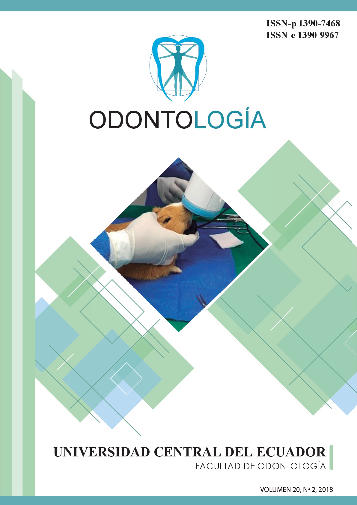
Histological and radiographical study of the alveolar post-extraction preservation with calcium sulfate and xenoinjerto in guinea pigs
Keywords:
Preservation, Alveolus, Alveolar ridge, Calcium Sulfat, XenograftAbstract
After performing an extraction, a process of bone remodeling begins, leaving as a consequence a decreased osseous volume that can prevent the placement of a dental implant in optimal conditions. Objective: To evaluate post-exodontia alveolar preservation using calcium sulfate or xenograft in guinea pigs compared to spontaneous healing. Materials and Methods: 30 male guinea pigs, four months old, assigned in three groups of 10 guinea pigs each were used: G1 Calcium sulphate, G2 Xenograft (bovine) G3 Negative control (without bone substitute). Exodontia of the lower right central incisor was curetted and washed with physiological serum, group G1 and G2 were filled with the corresponding biomaterial, group G3 did not receive any biomaterial. The suture was point in X with Vicryl (4/0). Standardized radiographs were taken in the immediate postoperative period and after 40 days, the alveolar crest was measured mesially, distally and coronally. At 40 days the guinea pigs were sacrificed, obtaining the segment of alveolar bone for the histological analysis of the alveolus. The data were analyzed through of the Kruskal Wallis test and chi square test with a level of significance of 5%. Results: The preservation of the alveolar crest was observed in mesial 2.92 (p = 0.025) and cervical 0.92 (p = 0.043). In the histological analysis, the spaces in the fibrous region in the middle part of the alveolus were 26.00 for calcium sulphate and 23.80 for xenograft (p = 0.011), apical 16.20 for calcium sulphate and 20 , 60 for the xenograft (p = 0.020), empty coronal spaces (p = 0.003), fibrous tissue (p = 0.010), bone regeneration (p = 0.019), hyaline areas (p = 0.010). Conclusion: Post-extraction alveoli are better preserved using Calcium Sulphate and Xenograft compared to spontaneous wound healing in guinea pigs.
Downloads
References
Ramirez K. Regeneración Ósea Guiada para preservación del Reborde Alveolar en la zona anterior. Revista Científica Odontológica. 2009; 5 (1): 29-33.
Leonida A, Todeschini G, Lomartire G, Cinci L, Pieri . Socket Preservation using Enzyme-treated Equine Bone Granules and an Equine Collagen Matrix: A Case Report with Histological and Histomorphometrical Assessment. The Journal of Contemporary Dental Practice. 2016; 17(11):890-896.
Salgado J, Zea del Río D, González J, Velosa J. Efectividad de las técnicas de preservación alveolar sobre alvéolos postexodoncia comparados con alvéolos sin preservar. Revisión sistemática de la literatura. Univ Odontol. Practica Clinica. 2014; 33(70):203-216.
Vargas L, Serrano C, Estrada J. Preservación de alvéolos postexodoncia mediante el uso de diferentes materiales de injerto. Revisión de la literatura. Univ Odontol. 2012; 31(66): 145-183.
Paolantonio M, Dolci M, Scarano A, D’ Archivio D, Di Placido G, Tumini V, Piattelli A. Immediate Implantation in fresh extraction socket. A controlled clinical and histological study in man. J. Periodontology. 2001; 72(11):1560-1571.
Guarinos J, Peñarrocha M, Sanchís J, Gay C, Sánchez M. La cresta alveolar atrófica en implantología oral. Anales de Odontoestomatología. 1995;119-130.
Carpenter J. Formulario de Animales Exóticos. In edición t, editor. Formulario de Animales Exóticos. Buenos Aires: Inter-médica; 2006:135-176.
Gómez Arcila V, Benedetti G, Castellar C, Fang L, Díaz A. Regeneración ósea guiada: nuevos avances en la terapéutica de los defectos óseos. Revista Cubana de Estomatología. 2014; 51(2):187-194.
Girano J. Exodoncia con conservación de reborde, injerto libre de paladar y plasma rico en fibrina. Reporte de caso. Revista ofcial de la Carrera Profesional de Estomatología.Facultad de Ciencias de la Salud – UPAGU. 2015;04(02):89-97
Jung-Chul Park, Ki- Tae Koo, Hyun- Chang Lim. The hidden X suture: a technical note on a novel suture technique for alveolar ridge preservation. Journal of Periodontal & Implant Science.. 2016; 46 (6):415-425.
Suárez D. Principios Básicos en Regeneración Ósea Guiada. Acta Bioclínica. 2012; 2(3):89-116.
Hoffmann O, Bartee B, Beaumont C, Kasaj A, Deli G, Zafiropoulos G. Alveolar Bone Preservation in Extraction Sockets Using Non-Resorbable dPTFE Membranes: A Retrospective Non-Randomized Study. Journal Periodontology. 2008; 79(8):1355-1369.
Arcesio R, Calvo J, Ramirez M, Maté J, Gómez G, Guardia J. Modelo experimental de la respuesta ósea a xenoinjertos de origen bovino. Estudio radiográfico e histomorfométrico. Acta Odontológica Colombiana 2011:1(2):12-26.
Araujo M, Lindhe J. Dimensional ridge alterations following tooth extraction. An experimental study in the dog. Journal of Clinical Periodontology. 2005; 32:212-218.
Piaggio L, Sacsaquispe S. Comparación histológica de la reparación ósea alveolar post-exodoncia utilizando una membrana colágena tipo esponja y un material de sulfato de calcio. Rev Estomatol Herediana. 2008; 18(2):93-98.
López M, Ayala M, Carbone C. Sulfato de Calcio en Regeneración ósea guiada. Acta Odontológica Venezolana. 2011; 49(4).
Rosales A, Franco J, Huacasi V, Larico B. Respuesta tisular a la aplicación de Brassica Rappa Campestris y Cestrun Parqui L`herit en alvéolos post- exodoncia de cobayos clase I. Revista Estomatológica del Altiplano. 2014; 1(2): 43-47.
Domínguez A, Torres C. Descripción histológica de la regeneración ósea en conejos implantados con hueso de bovino liofilizado (NUKBONE®). Investigación Universitaria Multidisciplinaria. 2006; 5(5):27-35.
Fickl S, Zuhr O, Whachtel H, Stappert CF, Stein JM, Hürzeler MB. Dimensional changes of the alveolar ridge contour after different socket preservation techniques. Journal of Clinical Periodontology. 2008; 35:906-913.
Fickl S, Schneider D, Zhur O, Hinze M, Ender A, Jung RE, Hürseler MB. Dimensional changes of the ridge contour after socket preservation and buccal overbuilding: an animal study. Journal of Clinical Periodontology. 2009; 36:442-448.
Johansen J, Gilhuus M. Repair of the post-extraction alveolus in the guinea pig a histological and autoradiographic study. The Department of Periodontology and the Department of Oral Surgery and Oral Medicine, Dental Faculty, University of Oslo, Norway. 1968 Noviembre; descargado 2014:250-258.
Saletta J, Rodríguez F, De la Plaza A. Actualización en Preservación de Cresta Alveolar. Revisión de la literatura. Cient. Dent.. 2014; 11(2):7-16.
Scheyer ET, Heard R, Janakievski J, Mandelaris G, Nevins ML, Pickering SR, et al. A randomized, controlled, multicentre clinical trial of post-extraction alveolar ridge preservation. J Clin Periodontol. 2016;43(12):1188-99
Parashis A, Kalaitzakis Ch, Tatakis D, Tosios K. Alveolar Ridge Preservation Using Xenogeneic Collagen Matrix and Bone Allograft. International Journal of Dentistry. 2014; (1):1-10.
Cardaropoli D, Tamagnone L, Roffredo A, Gaveglio L, Cardaropoli G. Socket preservation using bovine bone mineral and collagen membrane: a randomized controlled clinical trial with histologic analysis. Int J Periodontics Restorative Dent. 2012;32(4):421-30.
Molina J, Marcuschamer G, Rumeu J, Santos A, Griffin T. Preservación del reborde alveolar. Porque y cuando. Periodoncia y Oseointegración. 2007; 17(4):229-237.
Cioban C, ZĂGĂNESCU R, Román A, Muste A, Beteg F, CÂMPIAN R, BOŞCA B. Early healing after ridge preservation with a new collagen matrix in dog extraction sockets: preliminary observations. Romanian Journal Morphology Embryology. 2013; 54(1):125-130.
Sims G. Un cartilago vascularizado y a predominancia de células en maxilar superior e inferior de cobayo (Cavia porcellus). Zbl. Vet. Med. C. Anat. Histol. Embryol. 1980; 10:52-60.
López J, Alarcón M. Sulfato de calcio: propiedades y aplicaciones clínicas. Rev. Clin. Periodoncia Implantol. Rehabil. Oral. 2011; 4(3):138-143.
Perelman-Karmon M, Kozlovsky A, Liloy R, Artzi Z. Socket site preservation using bovine bone mineral with and without a bioresorbable collagen membrane. Int J Periodontics Restorative Dent. 2012;32(4):459-65
Joshi C, Dani N, Khedkar S. Alveolar ridge preservation using autogenous tooth graft versus beta-tricalcium phosphate alloplast: A randomized, controlled, prospective, clinical pilot study. Indian Society of Periodontology. 2016; 20(4):429-434.
DuPont L. Atlas of dental radiography in dogs and cats : a practical guide to techniques and interpretation. firts edition ed. Winkel A, editor. Philadelphia, PA, USA: Elsevier’s Health Sciences Rights Department; 2009.
Troiano G, Zhurakivska K, Lo Muzio L, Laino L, Cicciù M, Russo L. Combination of Bone Graft and Resorbable Membrane for Alveolar Ridge Preservation: a Systematic Review, Meta-analysis and Trial Sequential Analysis2017. 1-17 p.