
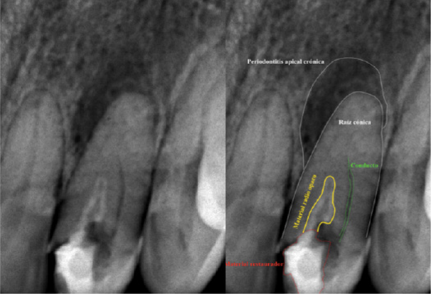
Upper left lateral incisor with anatomical variation.
DOI:
https://doi.org/10.29166/odontologia.vol24.n2.2022-e3927Keywords:
Anatomy, Root Canals, Conical ToothAbstract
This case report shows the need for internal and external anatomical knowledge of the dental organs to be treated in Endodontics, knowledge that must always be updated since there are unusual cases that require greater performance by the specialist to treat the case successfully. An endodontic retreatment was performed using 25 mm Triple Flex Kerr USA pre series #8 instruments, 25 mm WaveOne Gold Glider Dentsplay Sirona and 25 mm WaveOne Gold Small and Primary Dentsplay Sirona, SX ProTaper Universal Dentsplay Sirona file and burs Gates Glidden #1 and #2 Dentsplay Sirona. The obturation was carried out in a three-dimensional way with Tagger's hybrid technique. By repeating the endodontic treatment, it is possible to identify the entrance to two root canals in a single root with subsequent instrumentation and filling. Not all teeth that require endodontics are the same, so the treatment must be personalized and thoroughly analyzed before being treated.Downloads
References
González JM. Anomalías y displasias dentarias de origen genético-hereditario. Universidad de Sevilla, Sevilla, España. Avances en Odontoestomatología, 2012;28(6).
Carmona Marín L, Diente cónico: presentación de dos casos. Rev. Méd. Risaralda. 2014;20 (2):125-128.
Espinal Botero G. Estudio retrospectivo de anomalías dentales y alteraciones óseas de maxilares en niños de cinco a catorce años de las clínicas de la Facultad de Odontología de la Universidad de Antioquia. Revista Facultad de Odontología, Universidad de Antioquia. 2009;21(1): segundo semestre.
Bernal Sánchez K. Anomalías dentarias de número y forma. Caso clínico. Rev. Medigraphic Mx. 2014 ene-abr;vl(1): 9-14.
Krasner P. Anatomy of the pulp-chamber floor. Journal of Endodontics. 2004 jan;30(1).
Cohen S. Hargreaves K. Vías de la Pulpa. Décima edición. Editorial Elsevier 2011.
Acosta Vigorou S. Anatomy of the pulp chamber floor of the permanent maxillary first molar. Journal of Endodontics. 1978 jul. 4(7).
Alapati S. Maxillary canine with two root canals. Med Princ Pract. 2006;15:74-76.
Morgan LF. An evaluation of the crown-down pressureless technique. Journal of Endodontic 1984 oct;10(10).
Ponce de León Del Bello T. Crown-down tip desing and shaping. Journal of Endodontic. 2003 aug. 29(8).
Roane JB. The «balanced force» concept for instrumentation of curved canals. Journal of Endodontic. 1985 may;11(5).
Friedman S. Endodontic retreatment. Case selection and technique. Part 1: criteria for case selection. Journal of Endodontic. 1986 jan;12(1).
Stabholz A. Endodontic retreatment. Case selection and technique. Part 2: treatment planning for retreatment. Journal of Endodontic. 1988 dec;14(12).
Cohenca N. Endodontic retreatment of unusually long maxillary central incisors. Journal of Endodontic. 1996 may;22(5).
Taine M. Influence of endodontic treatment and retreatment on the fatigue failure load, numbers of cycles for failure, and survival rates of human canine teeth. Journal of Endodontic. 2017 dec;43(12).
Published
How to Cite
Issue
Section
License

This work is licensed under a Creative Commons Attribution-NonCommercial-NoDerivatives 4.0 International License.

