
Compact bone osteoma in the upper maxilla.
DOI:
https://doi.org/10.29166/odontologia.vol24.n2.2022-e3929Keywords:
Osteoma, Oral Cavity, Bone NeoplasmAbstract
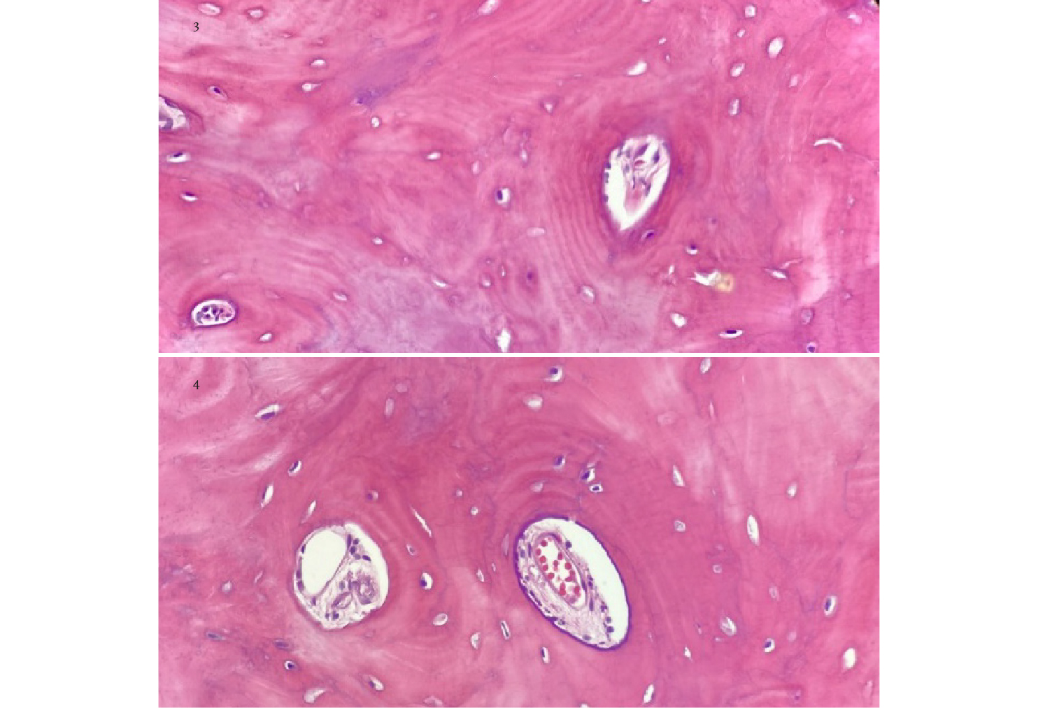
Osteoma is a benign lesion characterized by the presence of both cortical and cancellous bone tissue. Osteomas show slow but continuous growth, and can be single or multiple lesions with variation in size. Case report: 54-year-old male patient who comes to the Odontis Dental Clinic presenting severe pain in the upper left region associated to molar area. Radiographic and tomographic analysis were performed and then surgical removal of the lesion and the corresponding histopathological study. The presence of dense and compact lamellar bone with systems similar to those of Havers was found in the biopsy; no mitotic or atypical cellular activity was seen. A peripheral osteoma of compact bone in the upper jaw is diagnosed in close relationship with the maxillary sinus.Downloads
References
Neville B. Oral and maxillofacial pathology. 4th ed. Saunders/Elsevier; 2017.
Woldenberg Y, Nash M, Bodner L. Peripheral osteoma of the maxillofacial region: diagnosis and management. A study of 14 cases. Med Oral Patol Oral Cir Bucal. 2005:E139-42.
Shakya H. Peripheral osteoma of the mandible. J Clin Imaging Sci. 2011:1-56.
Colletti G, Autelitano L, Rabbiosi D, Tewfik K, Frigerio A, Biglioli F. Parosteal osteoma arising in an iliac bone graft used for mandibular reconstruction. J Oral Maxillofac Surg. 2012;70:477-80.
Boffano P, Bosco G, Gerbino G. The surgical management of oral and maxillofacial manifestations of Gardner syndrome. J Oral Maxillofac Surg. 2010;68:2549-54.
Lew D, DeWitt A, Hicks R, Cavalcanti M. Osteomas of the condyle associated with Gardner’s syndrome causing limited mandibular movement. J Oral Maxillofac Surg. 1999;57:1004-9.
Rappaport JM, Attia EL. Pneumocephalus in frontal sinus osteoma: A case report. J. Otolaryngol. 1994;23:430-436.
Bodner L, Gatot A, Sion-Vardy N, Fliss DM. Peripheral osteoma of the mandibular ascending ramus. J. Oral Maxillofac. Surg. 1998;56:1446-1449.
Halawi AM, Maley J, Robinson R, Swenson C, Graham S. Craniofacial osteoma: Clinical presentation and patterns of growth. Am. J. Rhinol. Allergy. 2013;27:128-133.
Larrea N, Valmaseda E, Berini L, Gay C. Osteomas of the craniofacial region. Review of 106 cases. J. Oral Pathol. Med. 2008;(37):38-42.
Societa’ Italiana di Chirurgia Maxillo-Facciale (sicmf). Trattato di Patologia Chirurgica Maxillo-Facciale Torino: Minerva Medica; 2007.
Ziccardi VB, Smith JA, Braun TW. Osteoma of the maxillary antrum. Oral Surg. Oral Med. Oral Pathol. Oral Radiol. Endod. 1995;(80):378-379.
Loukas M, Hulsberg P, Tubbs RS, Kapos T, Wartmann CT, Shaffer K, et al. The tori of the mouth and ear: A review. Clin. Anat. 2013;(26):953-960.
Yamasoba T, Harada T, Okuno T, Nomura Y. Osteoma of the middle ear: Report of a case. 1990;(116):1214-1216.
Sayan NB, Üçok C, Karasu HA, Günhan Ö. Peripheral osteoma of the oral and maxillofacial region: A study of 35 new cases. J. Oral Maxillofac. Surg. 2002; 60:1299-1301.
Espinosa FJ y cols. Osteoma mandibular periférico. Revista Mexicana de Cirugía Bucal y Maxilofacial 2017;13(2):60-64
Licéaga C, Del Bosque J, Aldape B, Montoya L, Morelos E, González V. Osteoma gigante en mandíbula. Reporte de Caso. 2013;23(4).
Prabhuji M, Kishore H, Sethna G, Moghe A. Peripheral osteoma of the hard palate. J Indian Soc Periodontol. 2012;16:134-7.
Seo-Young A, Chang-Hyeon A, Karp-Shik C. Giant osteoma of the mandible causing brething problem. Korean Journal of Oral and Maxillofacial Radiology. 2006; 36:217-220.
Published
How to Cite
Issue
Section
License

This work is licensed under a Creative Commons Attribution-NonCommercial-NoDerivatives 4.0 International License.