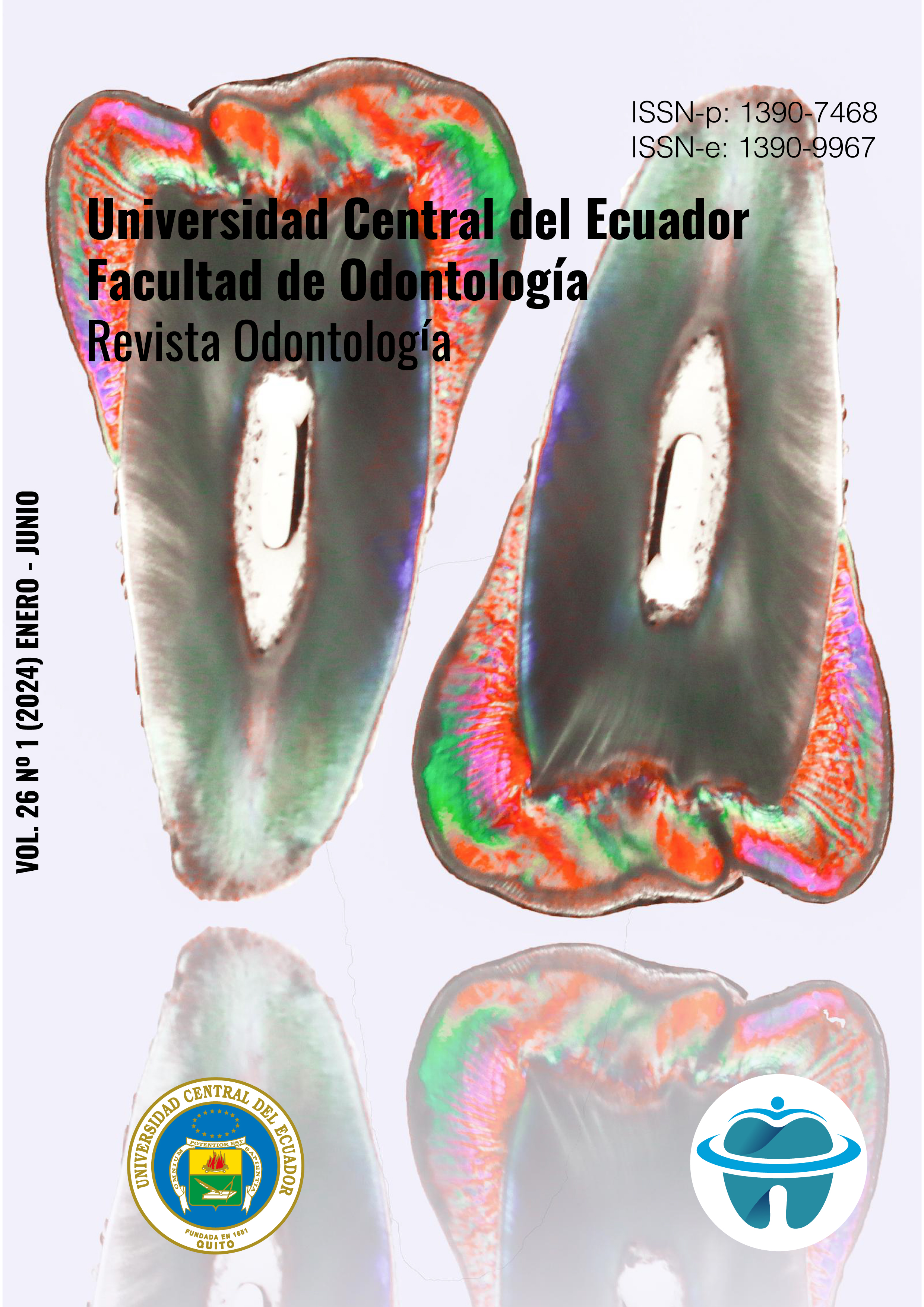
Dens invaginatus an esthetic and functional anomaly: findings on tomography of an ecuadorian population
DOI:
https://doi.org/10.29166/odontologia.vol26.n1.2024-e5887Keywords:
Dens in Dente, incisors, computed tomography, permanent dentition, dental anomalyAbstract
Among the structural development anomalies of a dental organ, dens invaginatus (dens in dente) is characterized by having an enamel invagination of varying depth, which could be associated with pulp, aesthetic and/or functional problems. Objective: Determine the prevalence of dens in dente in maxillary incisors according to sex, bilaterality and classification. Materials and methods: Cross-sectional observational study in 355 digital tomography scans from the period 2018-2021. Where 1420 maxillary incisors were analyzed in different cuts (axial, sagittal and coronal) to identify and classify the dens invaginatus. Statistical tests were carried out for the comparison of proportions with the help of the Z test and the association of variables with the Chi Square test (p < 0.05). Results: The prevalence of dens in dente was observed in 180 maxillary incisors (13%) present in 110 tomographies. 94% of cases corresponded to the maxillary lateral incisor, according to sex, no association was found with the presence of the dens invaginatus and the bilateral condition was expressed in 60.9%. Oehlers type I occurred in 97% of all cases and 3% for type II. Conclusions: Most cases of dens invaginatus were found in maxillary lateral incisors.
Downloads
References
Mabrouk R, Berrezouga L, Frih N. The Accuracy of CBCT in the Detection of Dens Invaginatus in a Tunisian Population. Int J Dent. 25 de enero de 2021;2021:8826204.
Różyło TK, Różyło-Kalinowska I, Piskórz M. Cone-beam computed tomography for assessment of dens invaginatus in the Polish population. Oral Radiol. 1 de mayo de 2018;34(2):136-42.
Chaturvedula BB, Muthukrishnan A, Bhuvaraghan A, Sandler J, Thiruvenkatachari B. Dens invaginatus: a review and orthodontic implications. Br Dent J. marzo de 2021;230(6):345-50.
Tomes J. A Course of Lectures on Dental Physiology and Surgery, Delivered at the Middlesex Hospital School. Am J Dent Sci. abril de 1848;8(3):209-46.
Varun K, Arora M, Pubreja L, Juneja R, Middha M. Prevalence of dens invaginatus and palatogingival groove in North India: A cone-beam computed tomography-based study. JCD. 5 de enero de 2022;25(3):306.
Alkadi M, Almohareb R, Mansour S, Mehanny M, Alsadhan R. Assessment of dens invaginatus and its characteristics in maxillary anterior teeth using cone-beam computed tomography. Sci Rep. 5 de octubre de 2021;11:19727.
Malmgren B, Thesleff I, Dahllöf G, Åström E, Tsilingaridis G. Abnormalities in Tooth Formation after Early Bisphosphonate Treatment in Children with Osteogenesis Imperfecta. Calcified Tissue International. 1 de agosto de 2021;109.
Riojas MT. Anatomía dental. 2a ed. México: El Manual Moderno; 2009.
Rickne C, Weiss G. Woefel, Anatomía dental. 8a ed. Philadelphia: Wolters Kluwer; 2012. 504 p.
Ash M, Nelson S. Wheeler, Anatomía Fisiología y Oclusión Dental. 8a ed. Madrid: Elsevier, Saunders; 2004.
Shafer WG, Hine MK. Tratado de patología bucal. Nueva Editorial Interamericana; 1988. 982 p.
Chen L, Li Y, Wang H. Investigation of dens invaginatus in a Chinese subpopulation using Cone-beam computed tomography. Oral Diseases. 2021;27(7):1755-60.
Yalcin TY, Bektaş Kayhan K, Yilmaz A, Göksel S, Ozcan İ, Helvacioglu Yigit D. Prevalence, classification and dental treatment requirements of dens invaginatus by cone-beam computed tomography. PeerJ. 5 de diciembre de 2022;10:e14450.
Capar ID, Ertas H, Arslan H, Tarim Ertas E. A Retrospective Comparative Study of Cone-beam Computed Tomography versus Rendered Panoramic Images in Identifying the Presence, Types, and Characteristics of Dens Invaginatus in a Turkish Population. JOE. abril de 2015;41(4):473-8.
Izaz S, Bolla N, Dasari B, Guntaka S. Endodontic Management of Calcified Oehler’s Type IIIb Dens Invaginatus in Permanent Maxillary Lateral Incisor Using Cone Beam Computed Tomography. J Dent (Shiraz). septiembre de 2018;19(3):243-7.
Kfir A, Flaisher Salem N, Natour L, Metzger Z, Sadan N, Elbahary S. Prevalence of dens invaginatus in young Israeli population and its association with clinical morphological features of maxillary incisors. Sci Rep. 13 de octubre de 2020;10:17131.
Published
How to Cite
Issue
Section
License
Copyright (c) 2024 Carla Angeles Pazmiño Flores, Erika Elizabeth Espinosa Torres, Rafael Guillermo Campanella Maldonado, María Soledad Peñaherrera Manosalvas, Esteban Velásquez Viteri

This work is licensed under a Creative Commons Attribution-NonCommercial-NoDerivatives 4.0 International License.


