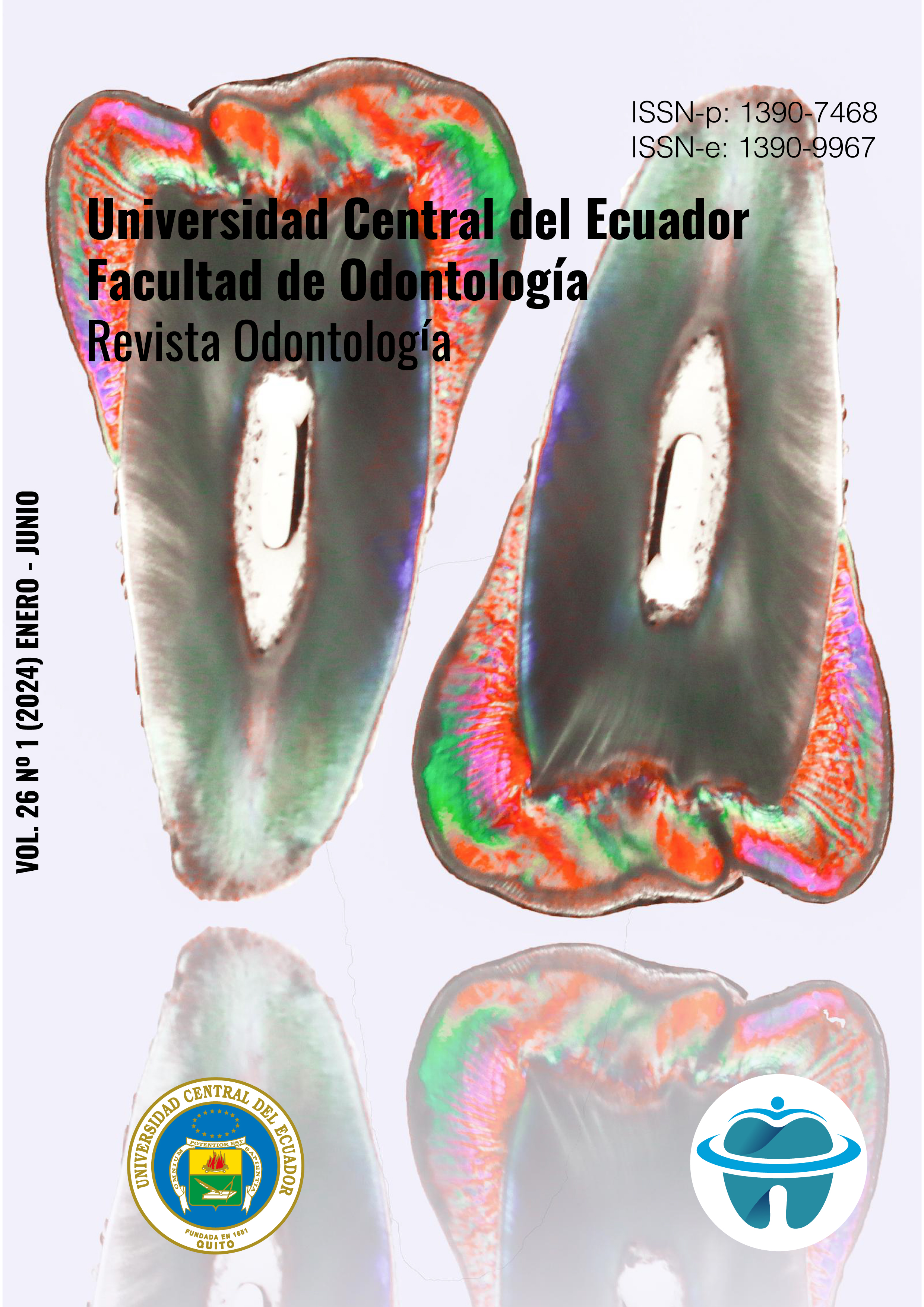
Cone Beam Tomography: unveiling the hidden dimensions of pulp chambers in maxillary and mandibular molars in Quito
DOI:
https://doi.org/10.29166/odontologia.vol26.n1.2024-e6055Keywords:
pulp chamber, molars, population, cone beam ct, endodonticsAbstract
Objetive. To evaluate the size of the pulp chamber in first and second maxillary and mandibular molars in the population of Quito, Ecuador, using cone beam computed tomography. Materials and Methods. The study included 100 maxillary and mandibular molar teeth from 52 tomographic scans of male and female patients aged between 15 and 30 years and between 31 and 46 years. Parameters measured included the distance from mesial pulp horn to mesial cusp center, distance from distal pulp horn to distal cusp center, distance between pulp horns, distance from the center of the pulp chamber floor to the furcation, and distance from the middle of the chamber from mesial to distal. Results. The distance between pulp horns and the middle of both distal and mesial cusps was greater in the first lower molars, and the mesio-distal distance of lower molars was greater for mandibular molars. Conclusions. The size of the pulp chamber in young patients is larger compared to adults due to the continuous deposition of secondary dentin throughout life. The measurement between the chamber floor and the furcation is not significantly representative, so equal care should be taken between patients of different ages during endodontic treatments.
Downloads
References
Azim AA, Azim KA, Deutsch AS, Huang GT. Acquisition of anatomic parameters concerning molar pulp chamber landmarks using cone-beam computed tomography. J Endod. 2014 Sep;40(9):1298-302. doi: 10.1016/j.joen.2014.04.002. Epub 2014 May 27. PMID: 25146010.
Leonardo, M. Endodoncia Tratamiento de Conductos Radiculares. Principios técnicos y biológicos vol.1 Sao Paulo Brasil. Artes Médicas Latimnoamérica 2005.
Canalda, C. Brau, E. Endodoncia Técnica Clínica y Bases Científicas. 3era edición. Barcelona España. ELSEVIER MASSON, 2014
Ge ZP, Ma RH, Li G, Zhang JZ, Ma XC. Age estimation based on pulp chamber volume of first molars from cone-beam computed tomography images. Forensic Sci Int. 2015 Aug;253:133.e1-7. doi: 10.1016/j.forsciint.2015.05.004. Epub 2015 May 14. PMID: 26031807.
Helmy MA, Osama M, Elhindawy MM, Mowafey B. Volume analysis of second molar pulp chamber using cone beam computed tomography for age estimation in Egyptian adults. J Forensic Odontostomatol. 2020 Dec 30;38(3):25-34. PMID: 33507164; PMCID: PMC8565657.
Maddalone, M., Citterio, C., Pellegatta, A., Gagliani, M., Karanxha, L., & Del Fabbro, M. (2020). Cone-beam computed tomography accuracy in pulp chamber size evaluation: An ex vivo study. Australian endodontic journal: the journal of the Australian Society of Endodontology Inc, 46(1), 88–93. https://doi.org/10.1111/aej.12378
Giongo, M., Gaona, P. & Victorino, F. (2016). Anatomical analysis of the pulp chamber of artificial teeth. RSBO Revista Sul-Brasileira de Odontologia, vol. 13(3), 194-198. https://www.redalyc.org/articulo.oa?id=153049441008
Sue, M., Oda, T., Sasaki, Y. et al. (2018). Age-related changes in the pulp chamber of maxillary and mandibular molars on cone-beam computed tomography images. Oral Radiol 34, 219–223. https://doi.org/10.1007/s11282-017-0300-1
Leonardo, M. Endodoncia Tratamiento de Conductos Radiculares. Principios técnicos y biológicos vol.1 Sao Paulo Brasil. Artes Médicas Latinoamérica 2005.
Ingle JI, Bakland LK. Endodoncia., Quinta Edición, editorial Mc Graw Hill, México, 2004; 2: 45-46, 409, 786.
Pagano J. Anatomía dentaria. Buenos Aires, Argentina: Mundi; 1965
Soares I. Goldberg. F. Endodoncia Técnica y Fundamento. Argentina. Editorial Médica Panamericana, 2003
Acosta SA. Vigouroux SA. Trugeda B. Anatomy of the pulp chamber floor of the permanent maxillary first molar. J Endod. 1978; 4: 214-9.
Figun ME. Anatomia odontológica funcional y aplicada. 2ª.ed. Buenos Aires: El Ateneo; 2001.
Allan, S. et al. (2004). Morphological Measurements of Anatomic Landmarks in Human Maxillary and Mandibular Molar Pulp Chambers. Journal of Endodontic 6(30), pp: 388 – 390.
Sue M, Oda T, Sasaki Y, & Ogura I. (2017). Age-related changes in the pulp chamber of maxillary and mandibular molars on cone-beam computed tomography images. Oral Radiology, 34(3), 219–223. https://doi.org/10.1007/s11282-017-0300-1
Azim A, Azim K, Deutsch A, & Huang G. (2014). Acquisition of Anatomic Parameters Concerning Molar Pulp Chamber Landmarks Using Cone-beam Computed Tomography. ournal of Endodontics, 40(9), 1298–1302. https://doi.org/10.1016/j.joen.2014.04.002
Adham A. Azim, Katharina A. Azim, Allan S. Deutsch, George T.-J. Huang. (2014) Acquisition of Anatomic Parameters Concerning Molar Pulp Chamber Landmarks Using Cone-beam Computed Tomography, Journal of Endodontics, Vol 40(9), 1298-1302, https://doi.org/10.1016/j.joen.2014.04.002. (https://www.sciencedirect.com/science/article/pii/S0099239914003689)
Supreet Jain, Ravleen Nagi, Minal Daga, Ashutosh Shandilya, Aastha Shukla, Abhinav Parakh, Afshan Laheji, Rahul Singh. (2017) Tooth coronal index and pulp/tooth ratio in dental age estimation on digital panoramic radiographs—A comparative study, Forensic Science International, Vol 277, 115-121, https://doi.org/10.1016/j.forsciint.2017.05.006. (https://www.sciencedirect.com/science/article/pii/S0379073817301779)
Published
How to Cite
Issue
Section
License

This work is licensed under a Creative Commons Attribution-NonCommercial-NoDerivatives 4.0 International License.


