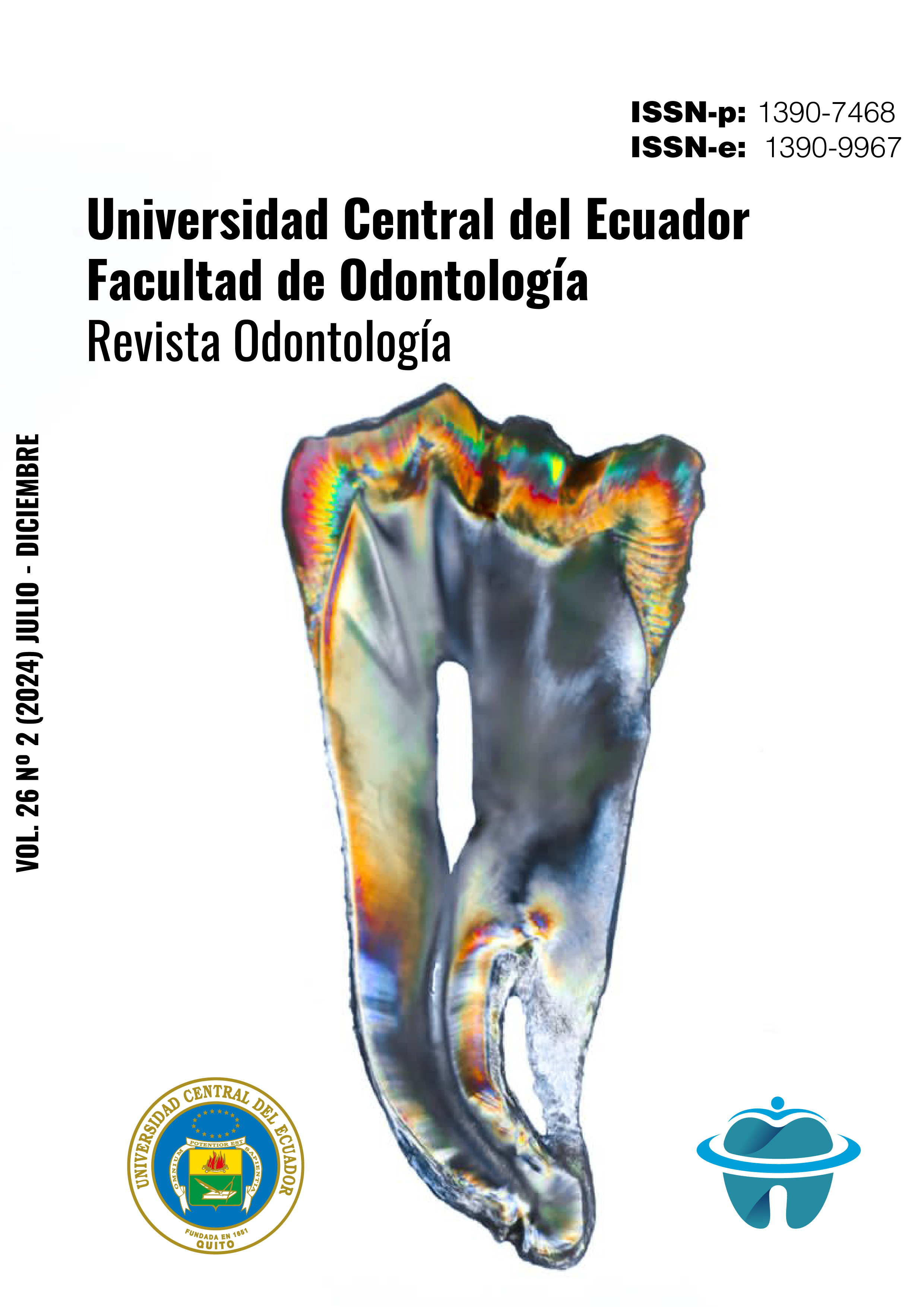
Mapping of the floor of the pulp chamber of upper first permanent molars using computed tomography
DOI:
https://doi.org/10.29166/odontologia.vol26.n2.2024-e6707Keywords:
dental pulp cavity, root canals, molars, computed axial tomographyAbstract
The relationship between the shape of the pulp floor determines the number of root canals found in a dental piece. The first investigations date back to 1978 by Sergio Acosta et al, as well as in 2004 by Krasner and Rankow; At present, this type of study helps to detect the canals found in the dental organ, even more so in the upper first molar, whose internal root anatomy is complex, especially to determine the presence of a second mesiopalatine canal (MB2). Aim: To determine by computerized axial tomography (CBCT) the main characteristics of the floor of the pulp chamber of the upper first molars. Materials and Methods: Descriptive and cross-sectional observational approach study in which 123 CBCTs were observed, the CBCT cohort was from the first to the last day of May 2023, to obtain a probabilistic sample of 107 CBCTs, in cut. axial at the cementoenamel limit. Results: Of the 187 upper molars examined, the most frequently found shape was the "Y" shape in 164 molars, of which between 3 and 4 canals were visualized, followed by the "four" shape observed in eleven teeth, as well as well as the "triangular" shape in six dental pieces, the "7" shape in four dental pieces and only the T and rectangular shapes were observed in 1 dental piece. Conclusions: The different anatomical features of the "rostrum canalium" in the upper first molars were determined by CBCT, the predominant pulp floor shape was the "Y" shape in dental organ 16.
Downloads
References
Battula, M, S., Kaushik, M., Mehra, N., & Singh, A. Endodontic management of maxillary first molar with unusual anatomy. Journal of conservative dentistry. 2022; 25(5), 569–572.
Krug, R., Connect, T., Beinicke, A., Solima,. S., Schubert, A., Kiefner, P., et. al. When and how do endodontic specialists use cone beam computed tomography?. Australian Endodontic Journal. 2019; 45(3): 365–372.
Barrett, M. The internal anatomy of the teeth with special reference to the pulp and its branches. Dent Cosmos. 2013; 67:581‑92.
Kyaw Moe, M., et al. Root Canal Configuration of Burmese (Myanmar) Maxillary First Molar: A Micro-Computed Tomography Study. International journal of dentistry, 2021, 3433343.
Pawar, A., Singh, S. New classification for pulp chamber floor anatomy of human molars. J Conserv Dent. 2020;23(5):430-435.
Acosta, S., Trugeda, S. Anatomy of the pulp chamber floor of the permanent maxillary first molar. J Endod. 1978 Jul;4(7), 214-9.
Versiani, M. The Root Canal Anatomy in Permanent Dentition. Alemania: Springer. 1st ed. 2019.
Paul Krasner DaHJRD. Anatomy of the Pulp-Chamber Floor. Journal of Endodontics. 2004; 30(1)
Castelucci, A. Endodontics. Edizioni Odontoiatriche, II Tridente. 2nd ed. 2009.
Pawar AM, Singh S. The morphology of the pulp chamber floor of permanent mandibular first and second molars in an Indian subpopulation-a descriptive cross-sectional study employing Pawar and Singh classification. PeerJ. 2022
Nelson, J. Wheeler. Anatomía, fisiología y oclusión dental. España: Elsevier Health Sciences Spain, 2015.
Published
How to Cite
Issue
Section
License
Copyright (c) 2024 Mónica Cristina Espín Míguez, Gustavo Morales Valladares, Erika Elizabeth Espinosa Torres

This work is licensed under a Creative Commons Attribution-NonCommercial-NoDerivatives 4.0 International License.


