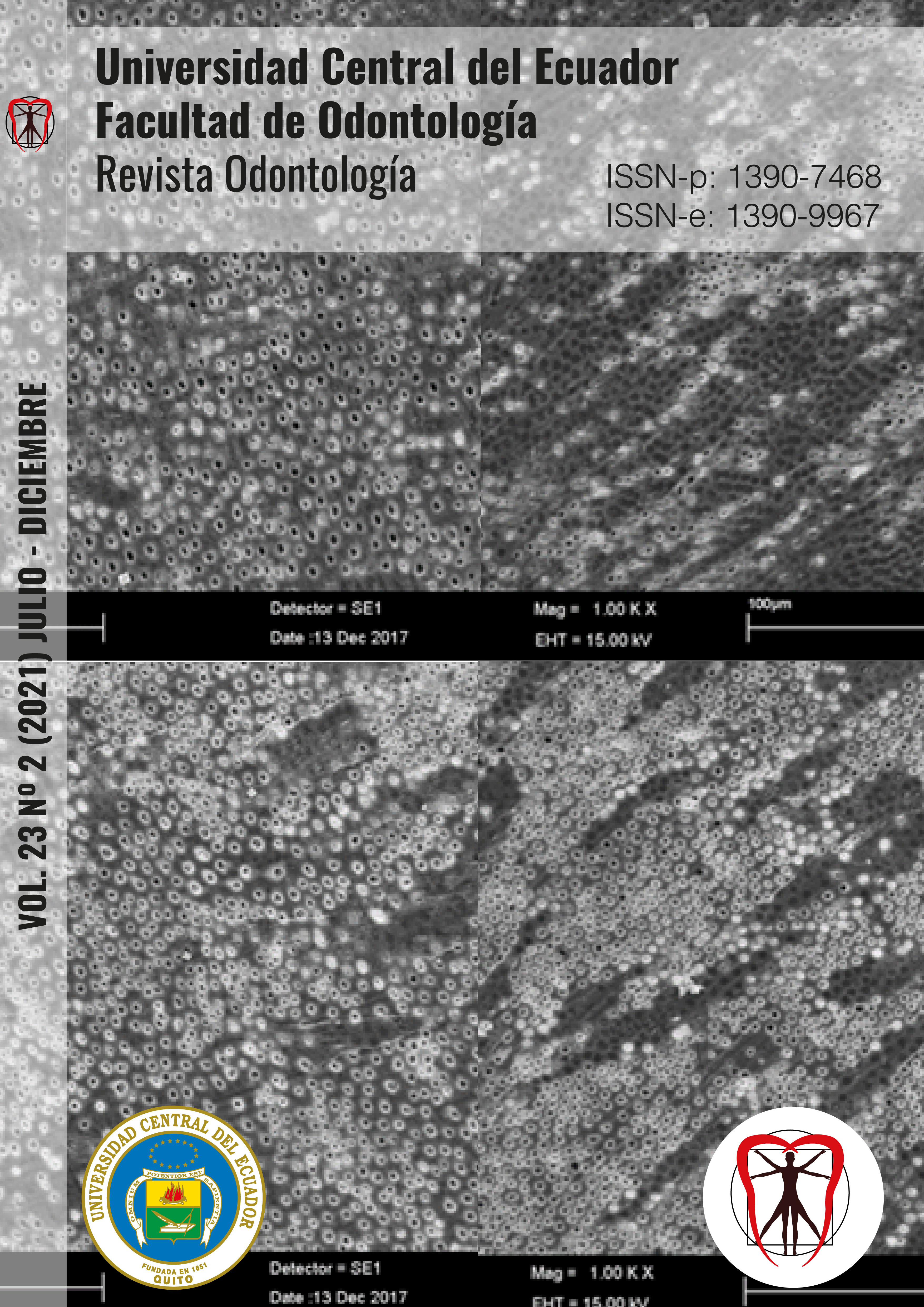
Conditioning, bonding, and cementation of orthodontic appliances in teeth with enamel alterations. Literature review
DOI:
https://doi.org/10.29166/odontologia.vol23.n2.2021-e3443Palabras clave:
Orthodontic bonding, adhesion enamel, amelogenesis imperfect, dental fluorosis, hypomineralization, enamel hypoplasiaResumen
Objective: Carry out a narrative review on the information available about the conditioning, bonding, and cementation of orthodontic appliances in teeth with enamel alterations. Materials and methods: Descriptive, retrospective research with a documentary design was carried out. 178 scientific articles were found in reliable sources such as Google Scholar, Scielo, PubMed, Scopus, Springer, Scientific Reports, and Elsevier related to the conditioning, bonding, and cementation of orthodontic appliances in teeth with enamel alterations; of which, 29 articles met the inclusion criteria and were selected. Results: Etching with 37% phosphoric acid plus composites remineralizing ingredients were more effective during orthodontic treatment in teeth with enamel alterations than other studied materials such as sodium hypochlorite, hydrochloric acid, bromelain gel, and papain gel. Conclusion: The materials that improve the conditioning, bonding, and cementation of orthodontic appliances in teeth with enamel alterations are phosphoric acid and sodium hypochlorite (NaOCI) with a concentration of 5.25%. Also, using deproteinizing agents could improve the resistance of the composite to eviction.
Descargas
Citas
García J. Patología y terapéutica dental: Operatoria dental y endodoncia. In García J. Patología y terapéutica dental: Operatoria dental y endodoncia. Barcelona, España: Elsevier; 2014. p. 1104. Available in: https://www.elsevier.com/books/patologia-y-terapeutica-dental/garcia-barbero/978-84-9022-655-1
Hinostroza M, Navarro R, Abal D, Perona G. Factores genéticos asociados a la hipomineralización incisivo-molar. Revisión de la literatura. Rev. Cient. Odontol. 2019; 7(1):148-156. DOI: 10.21142/2523-2754-0701-2019-148-156
Fleites Y, González K, Rico AM, Pacheco M, Del Toro L. Prevalencia de los defectos del desarrollo del esmalte en la dentición permanente. Medicentro Electrónica 2019; 23(3):177-191. Available in: http://scielo.sld.cu/scielo.php?script=sci_arttext&pid=S1029-30432019000300177
Reyes- Gasga J. Observación del esmalte dental humano con microscopia electrónica. Rev Tamé. 2013; 1(3):90-96. Available in: http://www.uan.edu.mx/d/a/publicaciones/revista_tame/numero_3/Tam133-06.pdf
Naranjo-Sierra MC. Terminología, clasificación y medición de los defectos en el desarrollo del esmalte. Revisión de literatura. Univ Odontol. 2013; 32(68):33-44.
Martín-González J, Sánchez-Domínguez B, Tarilonte-Delgado ML, Castellanos-Cosano L, Llamas-Carreras JM, López-Frías FJ, et al. Anomalías y displasias dentarias de origen genético-hereditario. Av Odontoestomatol. 2012; 28(6): 287-301. Available in: https://scielo.isciii.es/scielo.php?script=sci_arttext&pid=S0213-12852012000600004
Osorio-Tovar J, Naranjo-Sierra M, Rodríguez-Godoy M. Prevalencia de defectos de desarrollo del esmalte en dentición temporal, en una población bogotana. Rev. Salud Pública. 2016 Dec; 18(6): 963-975. DOI: http://dx.doi.org/10.15446/rsap.v18n6.48090
Acosta de Camargo MG, Natera A. Nivel de conocimiento de defectos de esmalte y su tratamiento entre odontopediatras. Rev. Odontopediatr. Latinoam. 2017;7(1). Available in: https://www.medigraphic.com/pdfs/alop/rol-2017/rol171d.pdf
Ulate J, Gudiño S. Hipomineralización incisivo molar, una condición clínica aún no descrita en la niñez costarricense.-ODOVTOS-Int. J. Dental Sc. 2015; 17(3):15-28. DOI: http://dx.doi.org/10.15517/ijds.v0i0.21482
Ramos L. Comparación de la superficie del esmalte dental post descementación de brackets metálicos después del acondicionamiento del esmalte con una sustancia remineralizante. Montevideo, Uruguay: Universidad de la República; 2010. Available in: https://repositorio.unal.edu.co/handle/unal/59092
Alfaro A, Castejón I, Magán R, Alfaro MJ. Molar-incisor hypomineralization syndrome. Rev Pediatr Aten Primaria. 2018; 20:183-188. Available in: https://pap.es/articulo/12651/molar-incisor-hypomineralization-syndrome
Ferreto-Gutiérrez I, Cáceres-Zapata H, Chan-Blanco JR. Comparación de la fuerza de adhesión de brackets a esmalte dental con un sistema exclusivo para ortodoncia y un sistema restaurativo. Revista Científica Odontológica. 2016; 12(2):8-14. Available in: https://www.redalyc.org/pdf/3242/324250005002.pdf
Bernales F. Evaluación de la aplicación de diferentes ácidos fosfóricos en la resistencia de unión de un adhesivo universal sobre el esmalte dental. Lima, Perú: Universidad Peruana Cayetano Heredia; 2019. Available in: https://repositorio.upch.edu.pe/handle/20.500.12866/6572
Aldred MJ, Savarirayan R, Crawford PJ. Amelogenesis imperfecta: a classification and catalogue for the 21st century. Oral Dis. 2003; 9(1):19-23. DOI: 10.1034/j.1601-0825.2003.00843.x
Pithon MM, Campos MS, Coqueiro Rda S. Effect of bromelain and papain gel on enamel deproteinisation before orthodontic bracket bonding. Aust Orthod J. 2016; 32(1):23-30. DOI: 10.2319/062911-423.1
Lang-Salas MG, Villarreal-Romero LA, Domínguez-Monreal JA, Cuevas‑González JC, Donohué‑Cornejo A, Reyes‑López SY, Zaragoza‑Contreras EA, Espinosa‑Cristóbal LF. Evaluación de la adhesión de sistemas adhesivos de grabado total en esmalte dental bovino usando un agente desproteinizante: un estudio in vitro. Rev ADM. 2020;77(1):22-27. DOI: 10.35366/OD201E
Romero S, Romero M, Natera A. Comparación de métodos para la remoción de resina residual posterior al descementado de aparatología fija de ortodoncia mediante el uso de gomas y discos. Odous Científica. 2018; 19(2): 9-21. Available in: http://servicio.bc.uc.edu.ve/odontologia/revista/vol19-n2/art01.pdf
Munive A, Cuellar MF. Protocolo de cementación indirecta de aparatología ortodóncica fija utilizando materiales de uso común. Rev ADM. 2019;76(6):315-321. Available in: https://www.medigraphic.com/cgi-bin/new/resumen.cgi?IDARTICULO=90448
Janiszewska-Olszowska J, Tandecka K, Szatkiewicz T, Stępień P, Sporniak-Tutak K, Grocholewicz K. Three-dimensional analysis of enamel surface alteration resulting from orthodontic clean-up -comparison of three different tools. BMC Oral Health. 2015; 15(1):146. DOI: 10.1186/s12903-015-0131-6
Santin GC, Palma-Dibb RG, Romano FL, de Oliveira HF, Nelson Filho P, de Queiroz AM. Physical and adhesive properties of dental enamel after radiotherapy and bonding of metal and ceramic brackets. Am J Orthod Dentofacial Orthop. 2015; 148(2):283-292. DOI: 10.1016/j.ajodo.2015.03.025
Gorucu-Coskuner H, Atik E, Taner T. Tooth color change due to different etching and debonding procedures. Angle Orthod. 2018; 88(6):779-784. Available in: https://doi.org/10.2319/122017-872.1
Krämer N, Bui Khac NN, Lücker S, Stachniss V, Frankenberger R. Bonding strategies for MIH-affected enamel and dentin. Dent Mater. 2018; 34(2):331-340. Available in: https://doi.org/10.1016/j.dental.2017.11.015
Ojeda A. Estudio comparativo de la efectividad de adhesión, entre la resina orthocem y heliosit orthodontic en el cementado de brackets y tubos metálicos en pacientes tratados en la clínica de postgrado de ortodoncia de la Facultad Piloto de Odontología de la Universidad de Guayaquil en el periodo 2013-2015. Universidad de Guayaquil. 2016. Available in: http://repositorio.ug.edu.ec/handle/redug/11602
Baherimoghadam T, Akbarian S, Rasouli R, Naseri N. Evaluation of enamel damages following orthodontic bracket debonding in fluorosed teeth bonded with adhesion promoter. Eur J Dent. 2016; 10(2):193-198. DOI: 10.4103/1305-7456.178296
Cruz-González A, Delgado-Mejía E. Experimental study of brackets adhesion with a novel enamel-protective material compared with conventional etching. The Saudi Dental Journal. 2019; 32. Available in: https://doi.org/10.1016/j.sdentj.2019.05.006
Trakinienė G, Petravičiūtė G, Smailienė D, Narbutaitė J, Armalaitė J, Lopatienė K, Šidlauskas A, Trakinis T. Impact of Fluorosis on the Tensile Bond Strength of Metal Brackets and the Prevalence of Enamel Microcracks. Sci Rep. 2019; 9(1):5957. Available in: https://www.nature.com/articles/s41598-019-42325-4
Nalcaci R, Temel B, Çokakoğlu S, Türkkahraman H, Üsümez S. Effects of laser etching on shear bond strengths of brackets bonded to fluorosed enamel. Niger J Clin Pract. 2017; 20(5):545-551. DOI: 10.4103/1119-3077.183245
Ferreira JTL, Borsatto MC, Saraiva MCP, Matsumoto MAN, Torres CP, Romano FL. Evaluation of Enamel Roughness in Vitro After Orthodontic Bracket Debonding Using Different Methods of Residua Estudio comparativo de la efectividad de adhesión,l Adhesive Removal. Turk J Orthod. 2020; 33(1):43-51. DOI: 10.5152/TurkJOrthod.2020.19016
Arnold WH, Haddad B, Schaper K, Hagemann K, Lippold C, Danesh G. Enamel surface alterations after repeated conditioning with HCl. Head Face Med. 2015;11:32. DOI: 10.1186/s13005-015-0089-2.
Gracco A, Lattuca M, Marchionni S, Siciliani G, Alessandri G. SEM‐Evaluation of enamel surfaces after orthodontic debonding: a 6 and 12‐month follow‐up in vivo study. Scanning. 37(5): 322-326 DOI: 10.1002/sca.21215
Zanini NA, Rabelo TF, Zamataro CB, Caramel-Juvino A, Ana PA, Zezell DM. Morphological, optical, and elemental analysis of dental enamel after debonding laminate veneer with Er,Cr:YSGG laser: A pilot study. Microsc Res Tech. 2021; 84(3):489-498. DOI: 10.1002/jemt.23605
Jablonski-Momeni A, Nothelfer R, Morawietz M, Kiesow Andreas, Korbmacher-Steiner H. Impact of self-assembling peptides in remineralisation of artificial early enamel lesions adjacent to orthodontic brackets. Scientific Reports. 2020; 10. Available in: https://doi.org/10.1038/s41598-020-72185-2
Özcan M, Sadiku M. Analysis of structural, morphological alterations, wettability characteristics and adhesion to enamel after various surface conditioning methods. Journal of Adhesion Science and Technology. 2016; 30:2453-2465. Available in: http://dx.doi.org/10.1080/01694243.2016.1184411
Ma Y, Zhang N, Weir MD, Bai Y, Xu HHK. Novel multifunctional dental cement to prevent enamel demineralization near orthodontic brackets. J Dent. 2017; 64:58-67. DOI: http://dx.doi.org/doi:10.1016/j.jdent.2017.06.004
Sharafeddin F, Safari M. Effect of Papain and Bromelain Enzymes on Shear Bond Strength of Composite to Superficial Dentin in Different Adhesive Systems. J Contemp Dent Pract. 2019; 20(9):1077-1081. Available in: https://pubmed.ncbi.nlm.nih.gov/31797833/
Hasija P, Sachdev V, Mathur S, Rath R. Deproteinizing Agents as an Effective Enamel Bond Enhancer-An in Vitro Study. J Clin Pediatr Dent. 2017; 41(4):280-283. DOI: 10.17796/1053-4628-41.4.280
Alkhudhairy F, Vohra F, Naseem M. Influence of Er,Cr:YSGG Laser Dentin Conditioning on the Bond Strength of Bioactive and Conventional Bulk-Fill Dental Restorative Material. Photobiomodul Photomed Laser Surg. 2020; 38(1):30-35. DOI: 10.1089/photob.2019.4661
Reymus M, Roos M, Eichberger M, Edelhoff D, Hickel R, Stawarczyk B. Bonding to new CAD/CAM resin composites: influence of air abrasion and conditioning agents as pretreatment strategy. Clin Oral Investig. 2019; 23(2): 529-538. Available in: https://doi.org/10.1007/s00784-018-2461-7
Zarif-Najafi H, Mousavi M, Nouri N, Torkan S. Evaluation of the effect of different surface conditioning methods on shear bond strength of metal brackets bonded to aged composite restorations. Int Orthod. 2019; 17(1):80-88. Available in: https://doi.org/10.1016/j.ortho.2019.01.009
Aglarci C, Demir N, Aksakalli S, Dilber E, Sozer OA, Kilic HS. Bond strengths of brackets bonded to enamel surfaces conditioned with femtosecond and Er:YAG laser systems. Lasers Med Sci. 2016; 31(6):1177-83. DOI: 10.100 Influence of Er,Cr: 7/s10103-016-1961-4
Jaâfoura S, Kikly A, Sahtout S, Trabelsi M, Kammoun D. Shear Bond Strength of Three Composite Resins to Fluorosed and Sound Dentine: In Vitro Study. Int J Dent. 2020; 2020: 4568568. Available in: https://doi.org/10.1155/2020/4568568
Khanal PP, Shrestha BK, Yadav R, Prasad Gupta DS. A Comparative Study on the Effect of Different Methods of Recycling Orthodontic Brackets on Shear Bond Strength. Int J Dent. 2021; 2021: 8844085. Available in: https://doi.org/10.1155/2021/8844085
Guerra A, Villacrés M. Comparación in vitro de la fuerza de adhesión sobre esmalte de brackets Clarity estándar (Transbond XT 3M) con los brackets Clarity APC Plus (3M), mediante una prueba de cizallamiento. OdontoInvestigación. 2015; 1(1). DOI: https://doi.org/10.18272/oi.v1i1.91
Descargas
Publicado
Cómo citar
Número
Sección
Licencia
Derechos de autor 2021 Catherine Andrea Vallejo Monrroy, Jackeline Beatriz Pedrosa Astudillo, Miriam Fernanda Ortega López, Lorenzo Puebla Ramos, Andrés Kenichi Noborikawa Kohatsu, Ronald Roossevelt Ramos Montiel

Esta obra está bajo una licencia internacional Creative Commons Atribución-NoComercial-SinDerivadas 4.0.


