
The root canal system of maxillary and mandibular second premolars using the dental diaphanization technique
DOI:
https://doi.org/10.29166/odontologia.vol26.n2.2023-e4392Keywords:
Endodontics, Tooth Root, dental pulp, root canalAbstract
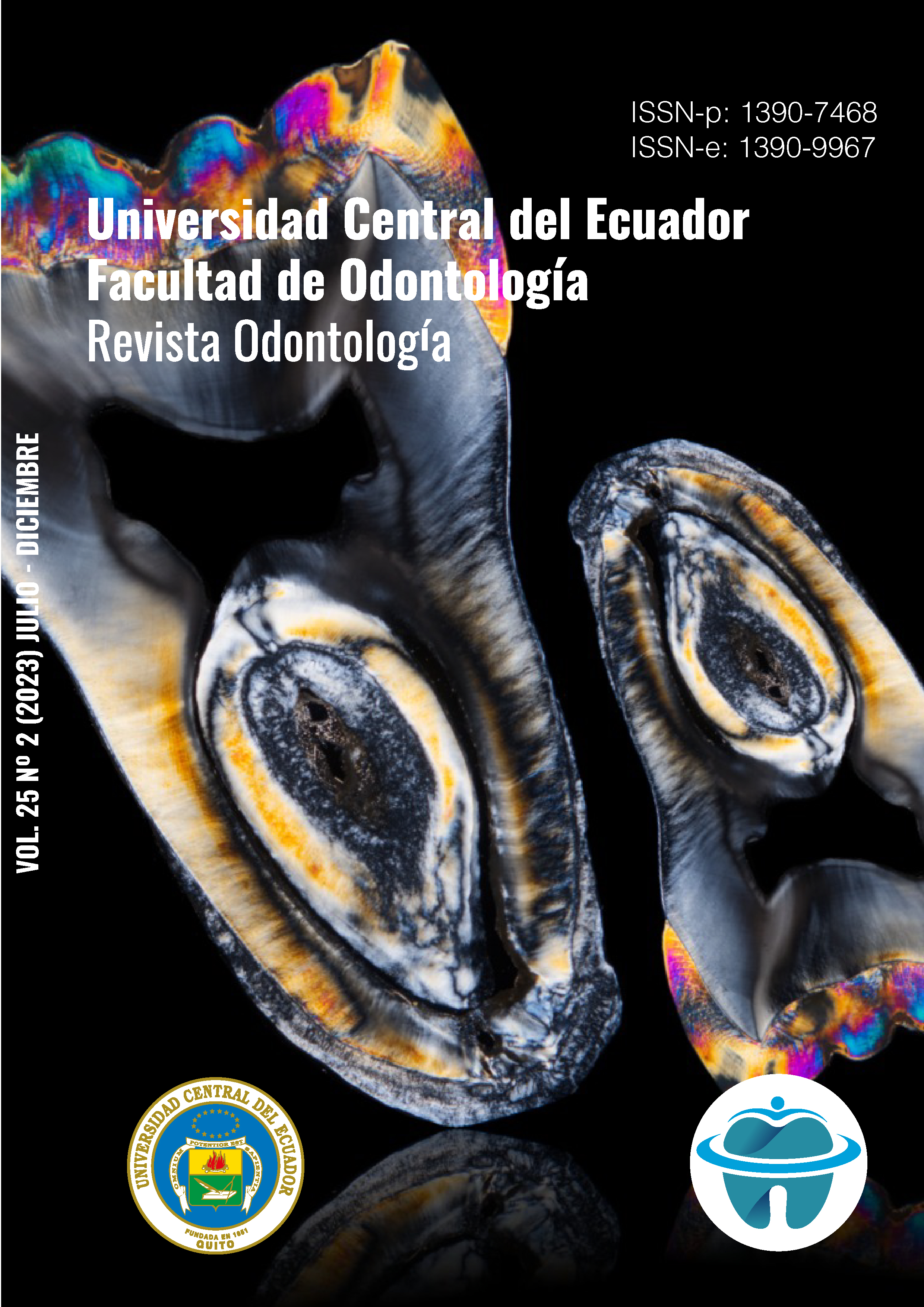
Objective: To identify the root canal system presenting the maxillary and mandibular second premolars, using the dental diaphanization technique. Materials and methods: 100 human second premolars were used, 50 upper and 50 lower, which were donated by owners of different dental centers in the city of Quito. They were sectioned at the cementoenamel level to dispose of the root. The root canal was permeabilized with a K # 10 file and Pelikan ® Indian ink was injected into the pulp chamber and root canals. To achieve decalcification, the teeth were immersed in a glass jar with 5% nitric acid. The dehydration process consisted of placing the teeth in 80%, 90% and 100% alcohol. Finally, the dehydrated teeth were stored in methyl salicylate for diaphanization, which was achieved in approximately two hours. Results: in the first 50 upper second premolars, the Vertucci type I classification was predominant with a total of n=25 (50%) pieces followed by the type IV classification n=16 (32%) type V n=6 (12 %) type III n=4 (4%) and classification type VII n=1 (2%). The remaining 50 lower second premolars presented the Vertucci type I classification, which was predominant with a total of n=37 (74%), followed by type IV n=8 (16%) and type III n=5 (10%).. Conclusion: Diaphanization is a technique of demineralization and whitening of extracted teeth, which allows direct and three-dimensional observation of their interior anatomy, allowing to know the variability of the root canal system of dental organs in a clear and didactic way.
Downloads
References
Rodríguez A. Endodoncia, Consideraciones Actuales. Primera Edición. Brasil. Editorial Amolca. 2003.
Riffo Muñoz, N. J., y Monardes Cortes, H. Determinación del número y topografía de los conductos radiculares en premolares inferiores. Universidad de Talca (Chile). Escuela de Odontología. 2008.
Favieri, R. A.; Rothier, A.; Fidel, R. Estudo da anatomía interna dos molares inferiores, submetidos ao processo de injeção por resina plástica. R.B.O. 1986; V. 43(6): 42-5
Greco, Y., García, J., Lozano, V. & Manzaranes, M. Morfología de los conductos radiculares de premolares superiores e inferiores. Endodoncia. 2009; 27 (1): 13-18. http://www.medlinedental.com/pdf-doc/ENDO/morfologia.pdf
Moreano Granizo, S. A. Técnica de diafanización dental. RECIMUNDO. 2019; 3(1): 724-741. https://doi.org/10.26820/recimundo/3.(1).enero.2019.724-741
Greco, Y., García, J., Bueno, R, Manzaranes, M. & Lozano, V. Técnicas de diafanización: estudio comparativo. Endodoncia. 2008; 26 (2): 85-92. http://diposit.ub.edu/dspace/bitstream/2445/67399/1/580905.pdf
Mashyakhy, M., Awawdeh, M., Abu-Melha, A., Alotaibi, B., AlTuwaijri, N., Alazzam, N., Almutairi, R., & Alessa, R. Anatomical Evaluation of Root and Root Canal Configuration of Permanent Maxillary Dentition in the Population of the Kingdom of Saudi Arabia. BioMed research international. 2022; 3428229. https://doi.org/10.1155/2022/3428229
Mageste Duque, Thais; Herrera Morante, Daniel Rodrigo; Randi Ferraz, Caio Cézar; Zaia, Alexandre Augusto; de Almeida, José Flávio Affonso; Figueiredo de Almeida Gomes, Brenda Paula Localización efectiva de un segundo conducto radicular en incisivos inferiores mediante magnificación, radiografía y diafanización. Revista Estomatológica Herediana. 2013; 23 (2): 57-62.
BRAVO, R., VALENZUELA, M.; CÁCERES, F. & SOTO, R. Aplicación de técnica de hidróxido de potasio y glicerina para diafanización dentaria. Int. J. Morphol. 2015; 33(2): 673-677.
Suh E, Karl E, Ramaswamy V, Kim-Berman H. The effectiveness of a 3D virtual tooth identification test as an assessment tool for a dental anatomy course. Eur J Dent Educ [Internet]. 2022 [citado el 16 de febrero de 2023];26(2):232–8. Disponible en: https://pubmed.ncbi.nlm.nih.gov/33982377/
Yang Y, Jiang C, Chen M, Zeng J, Wu B. Vertucci’s root canal configuration of 11,376 mandibular anteriors and its relationship with distolingual roots in mandibular first molars in a Cantonese population: a cone-beam computed tomography study. BMC Oral Health [Internet]. 2022 [citado el 16 de febrero de 2023];22(1):130. Disponible en: https://pubmed.ncbi.nlm.nih.gov/35429982/
Shrestha R, Srii R, Shrestha D. Diversity of root canal morphology in mandibular first premolar. Kathmandu Univ Med J (KUMJ). 2019;17(67):223–8.
Parekh V, Shah N, Joshi H. Root canal morphology and variations of mandibular premolars by clearing technique: an in vitro study. J Contemp Dent Pract [Internet]. 2011;12(4):318–21. Disponible en: http://dx.doi.org/10.5005/jp-journals-10024-1052
Vaillard Jiménez, Esther, Huitzil Muñoz, Enrique, & Castillo Domínguez, Loida. 2015).Características de los Canales Radiculares de Molares Temporales. International journal of odontostomatology, 9(1), 159-164. https://dx.doi.org/10.4067/S0718-381X2015000100024
Karobari, M. (2021). Root and Root Canal Morphology Classification Systems. Int J Dent. 6682189. doi: 10.1155/2021/6682189.
Wolf, T. G., Anderegg, A. L., Wierichs, R. J., & Campus, G. (2021). Root canal morphology of the mandibular second premolar: a systematic review and meta-analysis. BMC oral health, 21(1), 309. https://doi.org/10.1186/s12903-021-01668-z
Published
How to Cite
Issue
Section
License
Copyright (c) 2023 Andrés Alexander Castillo Chacón, Yessenia Alejandra Lozada Yundun, Gladys Rocío Hernández Santander, WWilliam Omar Granda Untuña, Michelle Estefania Aguayza Castro

This work is licensed under a Creative Commons Attribution-NonCommercial-NoDerivatives 4.0 International License.

